
Currently, the causes of the fibrous uterus are being studied; among the suspected factors are: hormonal abnormalities, familiarity with the development of uterine fibroids, obesity and an altered sensitivity of the uterus to growth factors.
The fibrous uterus can be a symptomatic or asymptomatic condition, depending on whether the uterine fibroids are large or not.
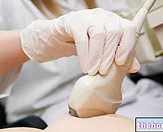
For the diagnosis of a fibromatous uterus, pelvic examination and pelvic ultrasound are essential.
Treatment of the fibrous uterus varies from patient to patient, based on the severity of symptoms, the patient's state of health and age.
Brief reminder of the uterus
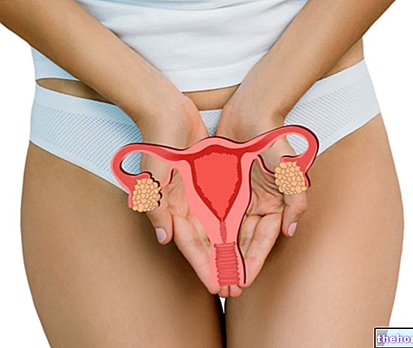
Learn and hollow, the uterus is the female genital organ, which serves to accommodate the fertilized egg cell (ie the future fetus) and to ensure its correct development, during the 9 months of pregnancy.
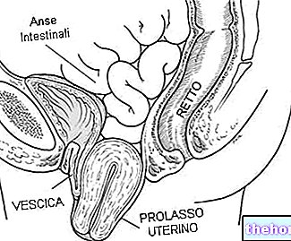
It resides in the small pelvis, precisely between the bladder (anteriorly), the rectum (posteriorly), the intestinal loops (above) and the vagina (below).
In the "span of life, the uterus changes its shape; if up to pre-pubertal age it has an elongated appearance similar to a glove finger, in adulthood it looks a lot like an inverted (or inverted) pear, while in the post-menopausal phase it gradually reduces its volume and becomes crushed.
From a macroscopic point of view, doctors divide the uterus into two distinct main regions: a more enlarged and voluminous portion, called the body of the uterus (or uterine body), and a smaller portion, called the neck of the uterus (or cervix).

What are Uterine Fibroids and why do they alter the Elasticity of the Uterus?
To understand why the fibrous uterus affects the elastic properties of the uterus, it is necessary to take a step back and review what uterine fibroids are.
Known as leiomyomas or uterine myomas, uterine fibroids are benign tumors of the uterus, which generally develop from the myometrium, which is the characteristic layer of muscle cells of the uterus.
Uterine fibroids appear as nodules of predominantly fibrous tissue, varying in size from a few millimeters to even 15-20 centimeters.
The fibrous uterus compromises the elasticity of the uterus, because the fibrous tissue of the uterine fibroids is a hard, inelastic and retracting tissue.
Did you know that ...
Uterine fibroids are very common; according to statistics, in fact, at least 80% of women can say, at the age of 50, that they have developed at least one uterine fibroid up to that moment.
How does the uterus become in women with fibromatous uterus?
In addition to compromising the elasticity of the uterus, the uterine fibroids that characterize the fibrous uterus make the affected organ more voluminous and thicker than normal; in pathology, a similar dimensional change in the uterus is called an enlarged uterus.
Types of uterine fibroid
Based on its location within the cell layers of the uterus, a uterine fibroid can be:
- Submucosal: is the type of uterine fibroid that tends towards the internal cavity of the uterus, ie towards the endometrium;
- Subserous: is the type of uterine fibroid that tends towards the external surface of the uterus;
- Intramural: it is the type of uterine fibroid that is kept inside the myometrium;
- Cervical: is the type of uterine fibroid that affects the neck of the uterus;
- Infralegamentary: it is the type of uterine fibroid interposed between the sheets of the so-called uterine ligament.
Scientific research has shown that uterine fibroids contain far more receptors for estrogen and progesterone than do normal uterine tissue.
This evidence has led scientists to hypothesize that the development of a uterine fibroid may depend on an abnormal concentration, in some parts of the uterus, of receptors for sex hormones;
Years of investigations relating to uterine fibroids have shown that women with a tendency to develop this type of benign tumors very often come from families in which consanguineous relatives (mothers, grandmothers, any sisters, etc.) have the same inclination.
This particularity has therefore led experts to believe that uterine fibroids and their possible consequences, such as for example the fibrous uterus, may have a genetic-family basis;
Several scientific studies have found that the lack of fine regulation of growth factors affects the development and growth of uterine fibroids;
Statistics show that uterine fibroids and fibrous uterus are more common in obese people.
Risk Factors: Who most often develops the Fibromatous Uterus?
According to statistics, conditions such as:
- Belonging to a family in which the problem of uterine fibroids and fibromatous uterus occurs among the female members;
- Membership of the black-skinned population;

- Obesity;
- Vitamin D deficiency;
- Consume large quantities of red meat and little fruit and vegetables;
- Abusing alcohol
- The early onset of menstruation.
The symptoms of the fibromatous uterus are non-specific manifestations, in the sense that they can characterize other pathologies affecting the uterus.

Complications
Women suffering from a severe form of fibromatous uterus (where by severe we mean that uterine fibroids are numerous and large) can experience complications, such as: reduced fertility and predisposition to spontaneous abortion, during pregnancy.
When to see a doctor?
A woman should always contact her trusted gynecologist whenever she suffers from one or more of the symptoms listed above; ailments such as pelvic pain, heavy menstruation, blood loss outside the menstruation period, etc., in fact, are all signs of something abnormal affecting the female reproductive system, uterus in particular.
pelvic; however, very often, doctors prescribe further investigations to patients with the disease in question, in order to investigate the severity of uterine fibroids and anatomical changes in the uterus.Additional investigations that a woman with a fibromatous uterus may need to carry out include:
- The transvaginal ultrasound;
- MRI of the pelvic organs;
- L "hysterosalpingography;
- The hysteroscopy.
FOCUSED ULTRASOUND GUIDED BY MAGNETIC RESONANCE
Focused ultrasound guided by magnetic resonance imaging is a completely innovative procedure, which allows to destroy uterine fibroids in a non-invasive way.
As can be guessed from its name, this procedure involves the use of ultrasounds and their addressing on the uterus, through a special instrument.
The big advantage of MRI guided focused ultrasound is that their use does not require surgical incisions.
What does "guided by magnetic resonance imaging" mean?
"Guided by magnetic resonance imaging" means that the attending physician uses the equipment for magnetic resonance imaging, to identify the precise point of the uterus on which to aim the ultrasound.
MINIMALLY INVASIVE SURGICAL PROCEDURES
Minimally invasive surgery implies: local anesthesia (rarely general), small surgical incisions, targeted action on the tumor or on the anomaly to be eliminated and fast recovery phase.
Indicated for patients still of childbearing age (therefore who may need to preserve the uterus), minimally invasive surgical procedures for the treatment of fibromatous uterus include:
- The embolization of the uterine artery. It is based on the principle of blocking the blood supply to the uterine fibroids, in order to induce their death due to the absence of oxygen and nutrients.
- Myolysis. It consists in exposing uterine fibroids to a laser or a beam of electric current, so as to cause the destruction of the benign tumor masses and the vessels that feed them.
- Laparoscopic or robotic myomectomy. Myomectomy is the surgery to remove uterine fibroids, leaving the uterus in place.
When performed with the laparoscopic or robotic technique, it means that the treating surgeon makes three small incisions on the abdomen (with the robotic technique, he uses a real robot to perform the elimination of uterine fibroids).

- Hysteroscopic myomectomy. It is myomectomy performed using the resectoscope, an instrument that emits electrical discharges capable of eliminating uterine fibroids.
The use of the resectoscope does not include incisions, but its insertion into the vagina and then into the uterus.
TRADITIONAL SURGICAL PROCEDURES
Traditional surgery implies: general anesthesia, large surgical incisions, little specific action on the target organ and long recovery times.
Indicated when the uterine fibroids are very large or when the patients are beyond the fertile age, the traditional surgical procedures for the treatment of the fibrous uterus consist of:
- Traditional myomectomy. It is the operation of myomectomy performed in laparotomy, that is through the incision and the opening of the abdomen.
- The hysterectomy. It is the surgery to remove the uterus; with its execution, the patient of childbearing age will no longer be able to have children.


.jpg)




-nelle-carni-di-maiale.jpg)


