What is Arthrocentesis?
Arthrocentesis is a commonly performed medical procedure for the diagnosis and treatment of certain joint diseases.

Indications of the arthrocentesis
Arthrocentesis is indicated for establishing a diagnosis, relieving symptoms, draining infected fluid, or infusing medications.
Diagnosis
- Joint effusion of unknown origin;
- Suspicion of septic arthritis;
- Crystal-induced arthropathy: gout and pseudogout;
- Hemorrhage (trauma);
- Rheumatoid arthritis, osteoarthritis and osteonecrosis.
- Reduction of intra-articular pressure and symptomatic relief of a large effusion (swelling);
- Limit joint damage from an infectious process by removing exudate from a joint.
- Diagnostic value. Arthrocentesis is mainly used in the diagnosis of gout, arthritis and infections. Synovial fluid can be tested for blood, pus, crystals, proteins and glucose, as well as cultured to determine the presence of pathogenic microorganisms. The appearance of the sample is also evaluated (color, viscosity, turbidity, volume of the synovial fluid, etc.) and the count of the cellular component (number of white or red blood cells) is performed. Each of these parameters can be useful in defining the cause of a particular pathology.
- The macroscopic aspect provides useful information on the degree of inflammation and the presence of haemarthrosis (collection of blood in a joint cavity).
- Crystal microscopy allows a precise diagnosis of gout (presence of monosodium urate crystals) and pseudogout (calcium crystal deposition disease pyrophosphate dihydrate).
- Microbiological studies are the key to confirming infectious conditions (example: Gram stain and microbiological culture for septic arthritis).
Diagnosis
Appearance
Viscosity
Particular results
Normal
Clear - yellow
High
Traumatic
Red
High
Presence of blood
Rheumatoid arthritis
Cloudy
Low
Haemagglutination on Rheuma test (or RA test)
Arthrosis
Clear - yellow
High (normal)
Possible presence of small fragments of cartilage
Gout
Cloudy
Decrease
Monosodium urate crystals (needle-like)
Pseudogout
Cloudy
Decrease
Calcium pyrophosphate (rhomboid) crystals
Septic arthritis
Low
Positive microbiological culture
Tuberculous arthritis
Cloudy
Low
Positive microbiological culture for acid-fast bacilli
- Therapeutic value: joint injection. Arthrocentesis can be useful for improving joint mobility and providing relief from pain and swelling. Drainage of joint effusion can also remove cells that participate in the inflammatory process; neutrophils and mononuclear leukocytes, in fact, release enzymes and chemicals that can damage sensitive tissues and induce joint degeneration. In order to quickly relieve joint inflammation and further reduce symptoms, a drug can be injected into the joint during arthrocentesis. This application is useful in the management of inflammatory conditions, such as rheumatoid arthritis, gout, tendonitis, bursitis and arthrosis.
The most frequently administered substances during arthrocentesis are:
- Corticosteroids: anti-inflammatories that act by reducing the symptoms of arthritis and limiting the accumulation of chemicals produced by the cells of the inflammatory process within the joint space. By attenuating the overactive immune system, corticosteroids can help reduce the " inflammation and to minimize tissue damage. Pain relief may last for a few months, but injections are not always effective and should not exceed four administrations per year for a specific joint.
- Hyaluronic acid: lubricates the joints, can be injected to improve mobility and relieve symptoms for periods of 6-12 months.
Procedure
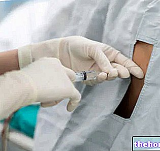
After having thoroughly cleaned the area to be treated, a liquid iodine solution is applied to the skin (example: Betadine ®), then a local anesthetic is injected into the area. The needle of a sterile syringe is inserted inside the joint, to easily collect synovial fluid by aspiration. After removing the excess fluid, the doctor may also inject a drug to treat some conditions. The needle is then removed and a dressing is applied over the entry site. Joints commonly undergoing arthrocentesis include: knee, shoulder, ankle, elbow, wrist, base of the thumb, and hand and foot joints. importance of this analysis, an ultrasound guide is sometimes advisable to facilitate arthrocentesis in difficult cases. Furthermore, ultrasound can be useful in revealing the presence of synovial fluid before aspiration and, subsequently, helping to distinguish some characteristic aspects of crystal-induced arthropathies.
Complications
Possible complications of arthrocentesis include bruising, minor local bleeding, and skin discoloration at the injection site. These effects are quite common. A rare but serious complication of arthrocentesis is infection (septic arthritis). joint corticosteroid medications, further complications can include atrophy and, if given too often, systemic side effects may occur.















-nelle-carni-di-maiale.jpg)
