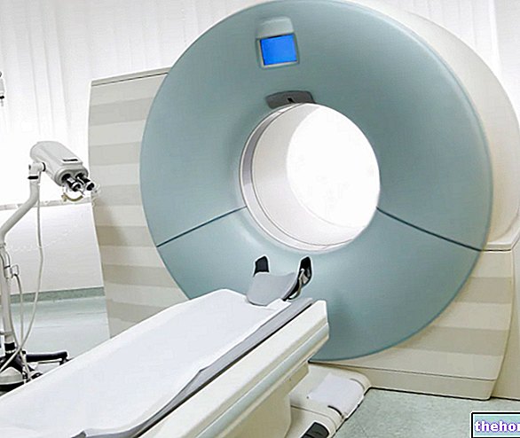
This nuclear medicine test uses radiopharmaceuticals or metabolic radio-compounds, ie substances normally present in the body, but marked with radionuclides capable of emitting corpuscular particles (positrons). A scanner (tomograph) detects the radiations emitted by the positrons of the tissue under examination and processes the collected data on the computer, returning mainly functional and metabolic information, useful for the diagnosis and orientation of the therapeutic protocol.
In clinical practice, the possible indications of PET are numerous. At present, the main fields of application can be identified in the field of neurological, cardiac and oncological diagnostics (diagnosis and follow-up of neoplasms, therapy monitoring, prognostic evaluation).
intravenously of a small amount of drugs and physiological agents labeled with radioactive isotopes (such as fluorine-deoxy-glucose F-18 or FDG F-18, i.e. glucose labeled with fluorine 18). In addition to "labeled glucose", other metabolic radio compounds used in positron emission tomography are methionine or dopamine. Once in circulation these radioactive tracers are distributed inside an organ or a specific biological tissue and emit particular particles, called positrons, which are captured by a special scanner (tomograph) and are translated into images that the nuclear medicine specialist interprets.
The tracers used in PET, such as, for example, Fluorine-18 (F-18) or "oxygen-15 (15-O), mimic the metabolic behavior of substances used by the body, ie glucose and oxygen from which they arise, accumulating where there is greater consumption (eg brain).This allows to differentiate each volume element of the organ under examination by oxygen or glucose consumption and to draw the diagnosis accordingly.
Learn more about the Basic Principle and How to Perform PET to get even more detailed images. This system allows to acquire PET and CT images in a single examination session with the consequent advantages:
- Reduction of examination times;
- Integrated diagnosis through the synergistic use of PET and CT information;
- Accurate interpretation of functional PET images based on anatomical CT images (anatomical-functional correlation);
- Improving the quality of functional PET images using CT anatomical information.

The images returned by positron emission tomography can therefore help to localize the presence of neoplastic processes in the body, highlighting the accumulation of this radiolabeled glucose analogue. Given the correlation highlighted between the high accumulation of this tracer and malignancy. of the tumor, PET has proved useful both in the diagnostic and prognostic fields, defining the site, the extent of the disease and the response to therapy of the cancer patient.
Therefore, the possibility of obtaining with PET information on the biological characteristics of the tumor, on the aggressiveness of the disease and on the presence of metastases is of considerable interest. This allows to correctly orient the choice of chemo and / or radiotherapy treatment, contributing to a more precise prognostic evaluation.

