Generality
The term vulva identifies the set of external genital organs of the woman.
In common parlance, the vulva is often referred to as the vagina, but the two terms CANNOT be considered synonymous. The vagina, in fact, is an internal musculo-membranous canal, which unites the uterus with the vulva. The latter, in addition to the external opening of the vagina, houses the meatus of the urethra, the clitoris and some small glands. bordered by two pairs of fleshy folds called vulvar lips.
The vulva performs various functions, such as the protection of the internal genitals, the perception of sexual pleasure during coitus and the sexual appeal for the man.
Synonyms
The vulva is also known as pudendum female or feminine pudendal. It is sometimes referred to as the vulvar complex.
Anatomy
The vulva is located between the inner faces of the thighs, in the central part of the perineum.
It extends from the hypogastric (suprapubic) region up to about 3 cm from the anus. It borders forward with the mount of Venus (see below) and posteriorly with the anus.
When the thighs are in contact, the vulva appears as a 10-12 cm long slit.
On the other hand, when the woman is in a gynecological position, with her thighs apart, the vulva has an ovoid shape with a vertical axis.
Structures of the vulva
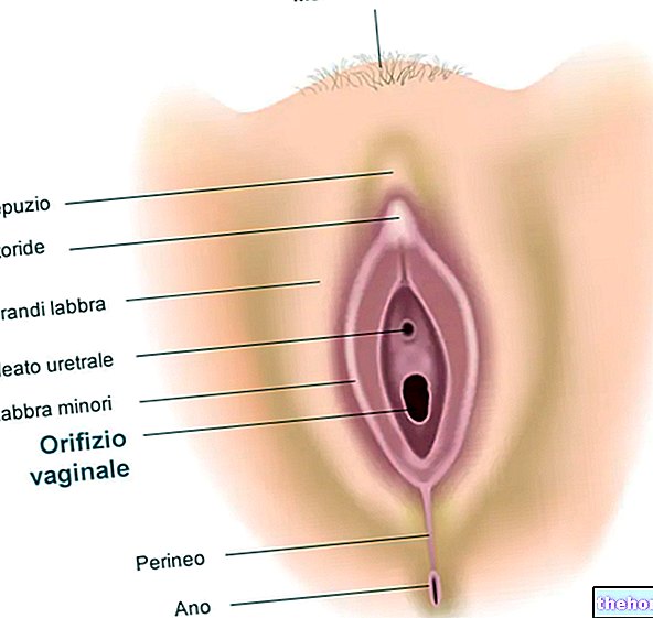
The female external genitalia - grouped under the single term vulva - include the following anatomical formations:
- Mount of Venus
- Big lips
- Small lips
- Erectile organs (clitoris and bulbs of the vestibule)
- Major (or Bartolini's) and minor (Skène's) vestibular glands
Mount of Venus
The mount of Venus (also known as the mount of the pubis) appears as a "raised skin area, due to the presence of abundant subcutaneous adipose tissue. It has a triangular shape, with the apex located at the bottom."
The mount of Venus is located above the pubic symphysis and overhangs the labia majora.
- Above it continues without a clear demarcation with the hypogastric region (lower part of the abdomen);
- laterally, on both sides, it is delimited by the folds of the groin;
- inferiorly it continues with the labia majora.
With its presence, the mount of Venus has the function of protecting the underlying bone, the pubic symphysis, during sexual intercourse.
The skin that covers it, hairless in childhood, is covered with pubic hair, long and sturdy, at the start of puberty; moreover, when sexual maturity is reached, the activity of the numerous sebaceous and sweat glands present in this area increases.
Big lips
The labia majora (or valve or labia majora) are two large skin folds of fibro-elastic connective tissue, rich in fat, parallel to each other and mutually welded, at the upper and lower extremities, in the vulvar commissures:
- superiorly, the labia majora start from the mount of Venus forming the anterior vulvar commissure (NB: the term commissure identifies the junction point between the two parts of a structure);
- posteriorly they join in the perineum, a couple of centimeters from the anus, forming the lower vulvar commissure (vulvar fork).
NB: between the vulvar fork and the vaginal orifice there is a small depression called navicular dimple.
Each large lip has two faces, one lateral (internal) and one medial (external); moreover, it has a free margin that delimits, with that of the large opposite lip, a median slit called the vulvar rim.
The medial aspect of the large lip adjoins the lateral aspect of the small ipsilateral lip (see below), through a depression called the interlabial sulcus.
The lateral face is separated from the inner face of the thigh by the genito-femoral or genito-crural sulcus.
In the adult woman, the labia majora measure on average 7-8 cm in length, 2-3 cm in width and 15-20 mm in thickness (in their middle part).
Normally more pigmented than the skin of the body, the labia majora are rich in sweat and sebaceous glands, whose secretion acts as a sexual attraction.
After puberty, the external (lateral) face of the labia majora appears covered with hair and becomes particularly active from the point of view of sebaceous secretion; moreover the skin that covers the valves becomes strongly pigmented and thicker. The inner face of the labia majora is instead covered with thin, pink and hairless skin.
After menopause, the labia majora become thinner, losing most of the fatty tissue; as a result, they appear thin, floppy and open.
The function of the labia majora is to protect the underlying structures, in particular the labia minora, the ostium (or vaginal meatus) and the external urethral orifice.
The labia majora are homologous to the male scrotum, with the difference that in the man the scrotal sac is joined medially longitudinally by a septum.

PLEASE NOTE: despite what is normally depicted in anatomical images, the vulvar rim is always closed at rest (it means that the labia majora completely hide the other structures of the vulva); to see the vulva "open" as shown in the figure, it is therefore necessary to spread the vulvar lips with the fingers.
During the sexual arousal phase, the labia majora tend to swell due to the increased blood flow; they also separate, making the labia minora more evident, which in turn increase in size and accentuate their color.
Little lips (or nymphs)
The labia minora (or nymphs or minor lips) are thin skin folds with a mucous, pinkish appearance. They are located inside the labia majora, from which they are separated by the nympholabial sulcus (or interlabial sulcus).
Anteriorly they split around the clitoris, forming the frenulum of the clitoris below it and above it a kind of semi-cylindrical envelope called hood or foreskin of the clitoris.
Unlike the labia majora, the labia minora typically do not join, but taper backwards and gradually disappear, merging into the inner part of the labia majora. More rarely, the lower ends of the labia minora meet in the midline, forming the frenulum of the labia minora.
The free margin of the labia minora is irregularly indented and floats freely.
In adult women, the length of the labia minora varies on average from 30 to 35mm, the width from 10 to 15mm and the thickness from 4 to 5 mm.
They have a pink, moist appearance and are hairless. Their conformation varies significantly with the constitutional characteristics; sometimes, for example, they are underdeveloped or even absent; at other times (like the labia majora) they can be doubled.
The sweat glands are missing in the labia minora, but the sebaceous glands abound, more numerous on the lateral side; some of these glands - called Fordyce granules - concentrate on the free edge of the labia minora, appearing as small, regular and uniform yellowish isolated papules.
During puberty the labia minora increasing in size, often protruding from the labia majora, from which they remained protected until the age of development.
In old age they thin and atrophy, taking on a dark brown color.
During sexual intercourse, the labia minora open and swell, thanks to the fibro-elastic structure, rich in nerve filaments and vessels, which characterizes them. Furthermore, the elastic component makes them easily expandable during sexual intercourse and at the moment of the passage of the fetus.
With their presence, the labia minora constitute the innermost protection of the urethral and vaginal orifices and of the clitoris. They are also marginally involved in the perception of sexual pleasure.
With their inner face, the labia minora delimit the vulvar vestibule.
Vulvar vestibule
The vulvar vestibule is a mucous region with a triangular shape, limited:
- forward and upward from the union of the labia minora around the clitoris (frenulum);
- laterally from the labia minora;
- back and down from the lower commissure or, if present, from the junction (frenulum) of the labia minora.
The following image, taken from wikipedia.org, highlights the vulvar vestibule delimiting it with a dotted line

The vestibule of the vagina houses:
- External urethral orifice (or urinary meatus): enclosed by the labia minora, it is located about 2 centimeters below the clitoris and allows urine to escape during urination. Close to it we find the outlet of the Skène glands (see below);
- Vaginal orifice: enclosed by the labia minora, it is located about 4 centimeters below the clitoris and allows the penis to enter the vagina, the menstrual flow to escape in childbearing age and, during childbirth, the passage of the fetus and fetal appendages. different women, the vaginal orifice differs in appearance depending on the presence of an intact hymen or its residues.
- Hymen: thin muscular-connective membrane which, in virgin women, occludes - usually partially - the vaginal orifice.
The hymen is separated from the labia minora by the nympho-hymenal sulcus or Hart's line.
The hymen can have different shapes from one woman to another (circular, half-moon, labiate, etc.). It normally tears (defloration) with the first sexual intercourse, causing a slight bleeding. The scar residues resulting from defloration are called hymenal lobules. After giving birth, these residues almost disappear (their remains constitute the hymenal caruncles).

Image taken from: https://en.wikipedia.org/wiki/Vulva
A detailed image of the vulva structures listed in the article is available at: http://www.britannica.com/media/full/633503/121148
Clitoris and Bulbs of the vestibule
The erectile apparatus of the female pudendal is formed by:
- a median organ, called the clitoris
- two lateral organs, called bulbs of the vestibule.
Clitoris
Also known as the female penis, the clitoris is an unequal erectile organ involved in a woman's sexual pleasure; it is in fact endowed with particular sensitivity, thanks to a rich network of sensitive vessels and nerve endings.
The clitoris is located at the top and front of the vulva, at the junction of the labia minora.
The clitoris can be divided into three parts:
- two lateral cavernous bodies, with an oblique orientation, also called roots; these are homologous cylindrical formations of the corpora cavernosa of the penis;
- the two roots converge medially and upwards to form, at the level of the pubic symphysis, a single and cylindrical organ called the body of the clitoris. This, for a short distance follows the direction of the roots, then abruptly bends forward (elbow or knee of the clitoris), and then goes down and back. Finally, it ends in a slightly swollen free formation with a blunt top, called the clitoral glans.
The clitoris is covered by a skin covering rich in sensitive nerve endings called the foreskin of the clitoris (just like the male foreskin covers the glans penis).
The clitoris hypertrophies (slightly increases in volume) when the woman is in a state of sexual arousal.
Clitoral hypertrophy (clitoromegaly) even at rest can occur in cases of hypertrichosis, polycystic ovary syndrome or abuse of anabolic steroids. However, it should be noted that there is no relationship between the size of the clitoris and the effectiveness of its function, although a higher level of androgens in women is normally associated with an increase in sexual desire.
Bulbs of the vestibule
They are two equal erectile formations (formed by cavernous tissue with large meshes), ovoid in shape, located at the base of the labia minora, developed on the lateral parts of the external openings of the urethra and vagina. They come into contact inferiorly with the Bartolini's glands and superiorly join under the elbow of the clitoris.
Attached glands
The glands attached to the vulva are divided into major (or Bartolini's) vestibular glands and minor vestibular glands.Bartholin's glands
The major vestibular glands or Bartholin's glands are two large glands located in the lower part of the labia majora, laterally and posterior to the vaginal orifice; their excretory duct opens through a small orifice in the nympho-hymenal sulcus, between the small lip and the orifice vaginal. They secrete a viscous liquid that participates in vaginal lubrication during sexual arousal.
In adult women, they have a variable volume from that of a pea to that of a small almond.
minor vestibular glands
Also called paraurethral or Skène glands, they are two small glands located in the vestibule, near the urethral meatus. They are considered the female analogue of the male prostate and, according to some researchers, would be the site of female ejaculation.


-cos-sintomi-cause-e-gravidanza.jpg)


