Edited by Dr. Giovanni Chetta
Fascial mechanoreceptors
Man represents the cybernetic system par excellence: 97% of decurrent motor fibers in the spinal cord are involved in the cybernetic process modality and only 3% are reserved for intentional activity (Galzigna, 1976). Cybernetics is the science of feed-back, the body must know moment by moment the environmental condition in order to be able to place itself instantly and appropriately for the purpose of carrying out the process. The sense can never be dissociated from motion: the environment must be continuously felt and evaluated, hence the need for gravity, synaesthesia, proprioception . "Being and functioning are inseparable" Morin; the reflection is the main road.
It is "the myofascial tissue that actually represents the largest sensory organ of our organism, it is from it that the central nervous system receives mostly afferent (sensory) nerves. The presence of mechanoreceptors, capable of causing effects at a local level and in general, it has been found abundantly in the fascia up to the visceral ligaments and in the cephalic and spinal dura mater (dural sac). We have seen that the organism reserves a great importance to the feed-back system. In fact, often in a mixed nerve the quantity of sensory fibers far exceeds the motor ones. What needs to be considered is that in the muscular innervation these sensory fibers derive only for about 25% from the well-known Golgi, Ruffini, Pacini and Paciniform receptors (type I and II fibers) while all the remainder originates from the "receptors" interstitial "(type III and IV fibers). These small receptors, which mostly originate as free nerve endings, as well as being the most numerous in our body are ubiquitous (their maximum concentration is in the periosteum) and therefore are present both in the muscular interstices than in the fascia. About 90% of them are demienized (type IV) while the rest have a thin myelin sheath (type III). The "interstitial" receptors have a "slower action than the type I and II receptors and in the past mostly nociceptors, thermo and chemoreceptors were considered. In reality, many of them are multimodal and most of them are mechanoreceptors that can be divided into two subgroups, based on their activation threshold by means of pressure stimuli: low-treshold (LTP) and high-treshold pressure (HTP) - Mitchell & Schmidt, 1977. L "activation, in certain pathological states of interstitial receptors sensitive to both painful and mechanical stimuli (mostly HTP) can generate painful syndromes in the absence of classic nerve irritations (eg root compression) - Chaitow & DeLany, 2000.
This sensory network, in addition to having an afferent sensing function of the positioning and movement of the body segments, influences, by means of intimate connections, the autonomic nervous system regarding functions, such as the regulation of blood pressure, heartbeat and breathing. tuning them, in a very precise way, to local tissue needs. The activation of the interstitial mechanoreceptors acts on the autonomic nervous system causing it to vary the local pressure of arterioles and capillaries present in the fascia, thus influencing the passage of plasma from the vessels to the extracellular matrix thus varying the local viscosity (Kruger, 1987). of interstitial receptors, as well as that of Ruffini receptors, is able to increase vagal tone by generating global changes at the neuromuscular, cortical and endocrine and emotional levels concerning a profound and beneficial relaxation (Schleip, 2003).
Deep manual pressures, performed statically or with slow movements, in addition to favoring the "gel to sol" transformation of the fundamental substance of the fascia (thanks to its thixotropic properties), stimulate Ruffini's mechanoreceptors (especially for tangential forces such as lateral stretching ) and a part of the interstitials inducing an increase in vagal activity with the relative effects on autonomous activities including a global relaxation of all muscles as well as mental (van denBerg & Cabri, 1999). The opposite result is obtained through strong and rapid manual skills that stimulate the corpuscles of Pacini and the Paciniforms (Eble 1960).
Myofibroblasts

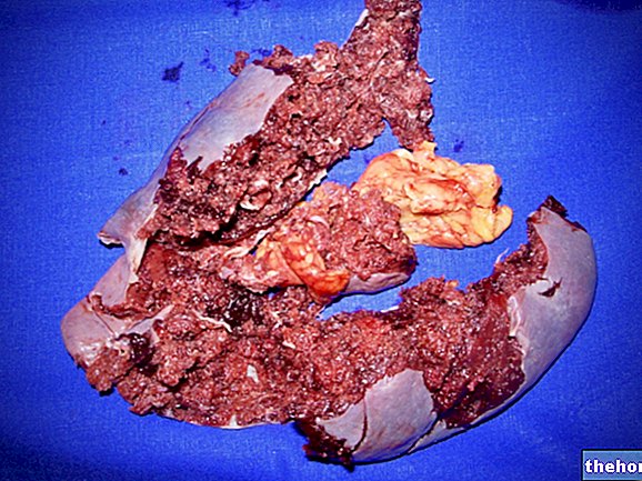
Given also the favorable configuration of the distribution of these contractile cells within the fascia, the probable role of these contractile structures is that of an accessory tension system such as to synergize muscle contraction providing an advantage in situations of danger for survival (fight and It is also very probable that through these smooth muscle fibers the autonomic nervous system, through intrafascial nerves, can "pre-tension" the fascia independent of muscle tone (Gabbiani, 2003, 2007). The presence of such cells in the covering capsules of the organs would explain e.g. how the spleen can shrink up to half its volume in a few minutes - a phenomenon observed in dogs in situations of strenuous effort in which the supply of the blood supply contained in it is required despite the capsular coating being rich in collagen fibers that allow only small variations in length - (Schleip, 2003).
Deep fascia biomechanics
From the biomechanical point of view, the thoraco-lumbar belt has the fundamental task of minimizing stress on the spine and optimizing locomotion.
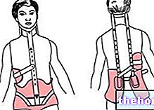

The erector muscles (multifidus) and intra-abdominal pressure, together with the psoas muscles, thus regulate lumbar lordosis three-dimensionally, thus assuming an important role as modulators of the transfer of forces between muscles and fascia.

There is no "universal optimal lordosis as it depends on the angle of flexion and the supported weight" (Gracovetsky, 1988).
Viscoelasticity of the fascia
As described, lifting heavy weights by putting the deep band under tension is the safest way to do it but it must also be done quickly in fact slowly it is possible to lift only ¼ of the weight that can be lifted at speed (Gracovetsky, 1988). This is due to the visco-elastic properties of the collagen fibers which determine an "elongation of the fascia when kept under tension for a long time. Because of its viscoelasticity, in fact, the fascia deforms under load in a short time, for this reason a continuous alternation of structures subjected to stress. The forces capable of elongating the fascia are greater the greater the state of tension already present (the more the fascia is elongated, the more difficult it will elongate further), in a non-linear manner (according to the studies of Kazarian of 1968, the response of collagen to the application of loads has at least two time constants: approx. 20 min and approx. 1/3 of a second) . The limit not to be exceeded in order to avoid breaking the fibers of the band is 2/3 of the maximum elongation. The "enemy" is therefore the splitting of the fascia from the periosteum; when the fascia is damaged, rehabilitation is very difficult, the subject presents a functional biomechanical and coordination imbalance. In children the fascia is immature, as the ossification of the vertebrae is incomplete, and thus the nerve impulses are not well transmitted. Consequently they move like people suffering from back pain caused by collagen damage forced to increase the "muscle activity (Gracovetsky, 1988).
The half-life period of collagen fibers in a non-traumatized tissue is 300-500 days, that of the "fundamental substance" (soluble portion of the ECM consisting of PGs / GAGs and specialized proteins) is 1.7-7 days (Cantu & Grodin 1992). Characteristics and arrangement of the new collagen fibers and of the fundamental substance also depend on the mechanical stress applied to the tissue.
Other articles on "Connective Band - Features and Functions"
- Connective tissue and Connective fascia
- Scoliosis - Causes and Consequences
- Scoliosis Diagnosis
- Prognosis of scoliosis
- Treatment of scoliosis
- Extra-Cellular Matrix - Structure and Functions
- Posture and tensegrity
- Man's motion and the importance of breech support
- Importance of correct breech and occlusal supports
- Idiopathic Scoliosis - Myths to Dispel
- Clinical case of Scoliosis and Therapeutic Protocol
- Treatment Results Clinical Case Scoliosis
- Scoliosis as a natural attitude - Bibliography


