Therapy includes rest from any heavy or dangerous activity for the ribs, applying ice to the painful area, and taking pain medication.

Anatomical recall on ribs and rib cage
The rib cage is the skeletal structure that serves to protect vital organs (such as the heart and lungs) and important blood vessels (aorta, hollow veins, etc.).
Located in the upper part of the human body, exactly between the neck and diaphragm, the rib cage includes:
- Posteriorly, 12 vertebrae;
- Latero-anteriorly, 12 pairs of ribs (or ribs);
- Anteriorly, the costal cartilages and a bone called the sternum.
Each pair of ribs originates from one of the 12 posterior vertebrae, which are part of the rib cage.
In the anterior part, the ribs end with the costal cartilages; the latter represent the point of union with the sternum only for the first 7 pairs of upper ribs. In fact, from the eighth to the tenth pair, the single ribs join (again through cartilage) to the upper rib (therefore the octaves to the seventh, the ninths to the octaves etc); while from the tenth to the twelfth pair, they are free.
Between the ribs are numerous muscles, known as the intercostal muscles. The intercostal muscles allow the rib cage to expand during breathing; therefore they play a fundamental role in the introduction of air into the lungs.

On particular occasions, the presence of one or more cracked ribs is also associated with an "alteration of the intercostal muscles.
The mechanism that leads to the appearance of a cracked rib is called, in specialist language, a rib crack.
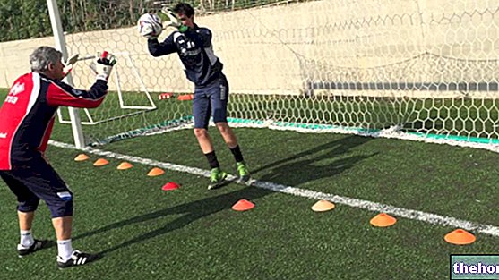
The rib crack is a fairly common injury of moderate severity.
and basketball.
PARTICULARITY OF PAIN
The pain that characterizes a cracked rib tends to get worse in some particular circumstances:
- When the patient breathes deeply.
- With compression of the injured chest area.
- With the twisting and bending movements of the body.
- When the patient sleeps on the side of the injured area.
If, due to severe pain, the patient finds it difficult to breathe normally, develops shortness of breath (dyspnoea), headache, dizziness, dizziness, recurrent tiredness / sleepiness and anxiety.
WHEN TO SEE THE DOCTOR?
When a blow to the chest causes persistent pain (especially during deep breaths), swelling and the formation of a hematoma, it is good to contact your doctor immediately or go to the nearest hospital for a deeper understanding of the situation.
Symptoms and signs for which it is good to contact your doctor.
- Presence of dyspnea
- Chest pain that increases rather than decreases
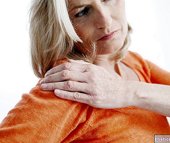
- Presence of pain in the shoulder or abdomen
- Cough and fever
COMPLICATIONS
Not being able to breathe deeply due to pain can have repercussions on lung health, in some cases very serious.
In fact, it can determine the onset of pneumonia and lung infections of various kinds.
Also, it's important to remind readers that a second blow to a cracked rib can cause it to break. A rib fracture is a far more serious injury than a crack, as the pointed ends of a broken rib can damage the thoracic blood vessels, lungs, heart, and major abdominal organs.
Please note: the fact that blood vessels and thoracic organs can be injured in the presence of a rib fracture distinguishes, from a purely medical point of view, the condition of a fractured rib from the condition of a cracked rib.
, swelling, etc.), and asks him about the symptoms:
- What do they consist of?
- Following what event did they appear?
- What movements or gestures heighten its intensity?
Questions of this kind allow us to broadly understand the basic problem and what caused it.
After the questionnaire, the physical examination ends with the palpation of the painful area (to see what the patient's response is), the auscultation of the lungs and heart (in search of any abnormal sounds) and the analysis of the head, neck , spinal cord and belly.
INSTRUMENTAL EXAMINATIONS
Instrumental radiological examinations are essential for achieving a correct and safe diagnosis.
Prescribed procedures usually include:
- X-rays. Generally, they can easily determine whether a rib is just cracked or fractured. They are unclear only in the presence of minor, non-distinct fractures.
X-rays are ionizing radiation that is harmful to health; however it should be remembered that the dose of such radiation is minimal. - The tomography. It provides a series of three-dimensional images, which very clearly reproduce the internal anatomy of the body.
It is very useful for excluding the presence of a fracture. In fact, it allows us to evaluate not only the bones of the rib cage, but also the health of the thoracic blood vessels, lungs and abdominal organs.
Its operation is based on the use of not negligible quantities of ionizing radiation. - Nuclear magnetic resonance (NMR). It is a radiological examination that provides for the patient to be exposed to completely harmless magnetic fields, without the need for harmful ionizing radiation.
Like the CT scan, it is useful for evaluating a wide range of elements: ribs, blood vessels passing through the chest, lungs and organs of the abdomen.

THE IMPORTANCE OF REDUCING PAIN
As anticipated, not being able to breathe deeply predisposes to the onset of pneumonia and lung infections.
Therefore, pain-relieving treatments have a "fundamental therapeutic importance: by reducing pain, they allow the patient to breathe deeply again, without suffering excessive discomfort.
Dos and don'ts to heal better
What to do:
- Keeping rest from any physical activity, be it a sport or a particularly heavy job. Failure to observe rest leads to a lengthening of recovery times.
- Take painkillers as recommended by your doctor. Their importance in preventing pneumonia and lung infections has already been discussed.
- Apply ice to the swollen and bruised area for at least 15-20 minutes 3-4 times a day.
- Breathe deeply or perform a controlled cough at least once or twice every hour. It serves to reduce the risk of pneumonia and lung infections.
What not to do:
- Do not wrap the chest with bandages. Otherwise, there is a risk of making deep breathing even more difficult.
- Not smoking.
- Do not make sudden movements or lift weights.
CURIOSITY €: THE THERAPY OF THE PAST
At one time, doctors treated cracked and fractured ribs by applying restraining bandages to patients' thorax to immobilize the affected area as much as possible.
With the discovery that this type of treatment increases the risk of pneumonia and lung infections, they immediately abandoned it and adopted the current method of treatment.









.jpg)


