Generality
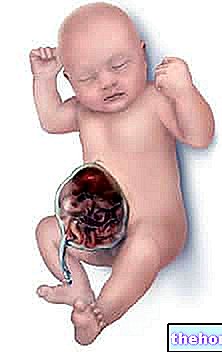
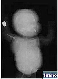
Hydrops fetalis is a serious medical condition characterized by the accumulation of fluid in the subcutaneous tissues and serous cavities of a fetus or newborn.
There are two subtypes of fetal hydrops: non-immune fetal hydrops and immune fetal hydrops.

Immune hydrops fetalis, on the other hand, almost always depends on a maternal-fetal blood incompatibility for the Rh factor.
The possible symptoms of fetal hydrops include: presence of fluid in the serous cavities or in the subcutaneous tissues (edema), respiratory problems, pallor, bruising, jaundice, anemia and heart failure.
The treatment of fetal hydrops depends on the triggering cause and the symptoms in progress.
What is fetal hydrops?
Hydrops fetalis is a serious medical condition characterized by the accumulation of fluid in at least two areas of the body of a fetus or newborn baby (newborn).
The accumulation of fluid can occur in the subcutaneous tissues - in these situations the doctors speak of edema - or in the serous cavities. The serous cavities usually affected by the fetal hydrops include:
- The abdomen. The accumulation of fluid in the abdominal cavity (or peritoneal cavity) is called ascites.
- The pericardium. The accumulation of fluid in the pericardial cavity is known as a pericardial effusion.
- The pleura. The accumulation of fluid in the pleural cavity is known as a pleural effusion.
Causes
Based on the triggering causes, doctors thought of distinguishing fetal hydrops into two subtypes: non-immune fetal hydrops and immune fetal hydrops.
NON-IMMUNE FETAL HYDROPE
Characterizing more than 90% of clinical cases, non-immune hydrops fetalis is the most common subtype of fetal hydrops.
Its presence is the result of an increase in interstitial fluids or a "lymphatic obstruction.
Causes of non-immune hydrops fetalis include:
- Cardiovascular conditions, such as arrhythmias, coronary embolism, the shunts arteriovenous, myocarditis, cardiac tumors, Fallot's tetralogy or Ebstein's malformation.
- Chromosomal abnormalities, such as Turner syndrome, trisomy 21 or Noonan syndrome.
- Infections of various kinds, such as toxoplasmosis, rubella, chicken pox, syphilis, Lyme disease, AIDS and infectious diseases caused by cytomegalovirus, herpes simplex virus, enterovirus or parvovirus.
- Pulmonary malformations, such as pulmonary hypoplasia.
- Malformations of the urinary tract.
- Episodes of congenital diaphragmatic hernia.
- A severe state of anemia, resulting from thalassemia or an iron deficiency.
IMMUNE FETAL HYDROPE
Immune fetal hydrops arises as a result of an incompatibility between the blood group of the mother and the blood group of the future unborn child. In fact, when the aforementioned situation arises, the mother produces antibodies to fetal erythrocytes, which attack the latter giving rise to various complications, including the accumulation of fluid in the subcutaneous tissues and / or in the serous cavities (ie the "hydrops fetalis).
In most cases, fetal immune hydrops results from a maternal-fetal incompatibility for the Rh factor (or Reshus factor).
Symptoms, signs and complications
The symptoms and signs of hydrops fetalis vary in relation to the severity of the hydrops fetalis itself. In other words, the milder forms of hydrops fetalis are responsible for less severe symptoms than the more severe forms.
LIGHT FORMS: TYPICAL SYMPTOMS
Generally, milder forms of fetal hydrops induce ascites and paleness.
Readers are reminded that, even in a mild form, fetal hydrops is still a serious medical condition.
SEVERE FORMS: TYPICAL SYMPTOMS
As a rule, the most severe forms of fetal hydrops cause:
- Breathing problems
- Appearance of cutaneous bruises or purple spots on the skin;
- Heart failure;
- Severe anemia;
- Severe jaundice;
- Edema in various parts of the body.
COMPLICATIONS
Hydrops fetalis is a highly lethal condition for affected children.
In cases of immune hydrops fetalis due to a "maternal-fetal incompatibility for the Rh factor, subjects who survive childbirth and even the following weeks are at high risk of kernicterus.
Kernitter is a particular form of encephalopathy, characterized by the accumulation of bilirubin in the brain tissue. Not surprisingly, kernitter is also known as bilirubin encephalopathy.
Diagnosis
Usually, for a correct diagnosis of fetal hydrops it is essential to resort to a "morphological prenatal ultrasound. In the images reported by the latter, there are typical signs of a" fetal hydrops:
- The presence of large amounts of amniotic fluid;
- The presence of a large placenta;
- The abnormal presence of fluid around certain organs of the fetus, including the liver, spleen, heart and / or lungs.
WHAT DIAGNOSTIC EXAMINATION ALLOWS TO ESTABLISH ITS SEVERITY?
To understand the severity of a "fetal hydrops, doctors may resort to an" amniocentesis or periodic morphological ultrasound scans.
HOW TO ESTABLISH THE SUBTYPE OF FETAL HYDROPE?
To determine the subtype of fetal hydrops present, doctors need to take a maternal blood sample and look for antibodies to fetal erythrocytes in it. The presence of these antibodies indicates that the fetal hydrops is of the immune subtype; their absence, on the other hand, means that the fetal hydrops is of the non-immune type.
Identifying the subtype of fetal hydrops is essential for planning the most appropriate therapy and hoping for a better (or less unfavorable) prognosis.
Therapy
The treatment of fetal hydrops varies in relation to several factors, including, mainly, the triggering causes and the symptoms in progress.
TREATMENT IN THE PRENATAL AGE
In prenatal age, fetal hydrops is treatable only in certain circumstances (eg presence of anemia at the origin of fetal hydrops).
In such circumstances, the typical treatment is an intrauterine fetal blood transfusion.
When the possibilities of prenatal treatment are lacking, doctors promote premature birth of the fetus, as the therapeutic possibilities for newborns are greater and more effective. Premature birth of the fetus can occur by means of particular drugs, which stimulate labor, or by caesarean section.
TREATMENT IN NEWBORN (OR POST-CHRISTMAS) AGE
The treatments of fetal hydrops in newborns include:
- The transfusion of blood, to clean the latter from the anti-fetal erythrocyte antibodies, passed from the mother to the child, when the first provided for the circulation of the blood in the second;
- The removal, by syringe, of the fluid accumulated in the pleural or abdominal cavities;
- Giving medicines to control heart problems (heart failure);
- The administration of drugs that stimulate the kidneys to eliminate excess fluid, present in the subcutaneous tissues;
- The use of breathing aids, such as machines for artificial ventilation.
Prognosis
Very often, fetal hydrops causes the baby to die either shortly before birth or shortly after birth. The prognosis, therefore, tends to be unfavorable.
Prevention
For several decades now, it has been possible to prevent fetal immune hydrops by means of a drug, called RhoGAM (or Rho immunoglobulin). Administered only and exclusively to pregnant women with a "maternal-fetal incompatibility for Rh factor, RhoGAM prevents the the mother's immune system produces antibodies to fetal erythrocytes, which are the agents triggering the condition of hydrope fetal immune.

