Generality
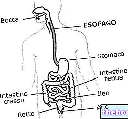
Esophageal diverticula are congenital or acquired sac-like extroversions of the esophagus wall, communicating with the lumen of the esophagus. In relation to the mechanism of formation, diverticula are distinguished by drive and traction. The former are due to the gradual ejection of the mucosa and submucosa. through an "area of weakness in the muscle wall, due to an increase in intraluminal pressure. The latter, on the other hand, occur as a consequence of traction forces exerted on the esophageal wall, due to an adjacent inflammatory process (example: involvement in inflammation of the mediastinal lymph nodes in tuberculosis). Esophageal diverticula are often asymptomatic, but are sometimes associated dysphagia and symptoms of varying severity related to esophageal dyskinesias.
Diagnosis
Physical examination results are often normal, as patients are frequently asymptomatic. However, many patients report episodes of dysphagia, chest pain, or food regurgitation.
Radiographic investigations of the esophagus or upper gastrointestinal tract allow to detect many of the non-symptomatic diverticula.
- Chest radiography and computed tomography allow identification of large esophageal diverticula, which may manifest as structures filled with air and / or fluid communicating with the esophagus.
- Fluoroscopy of the esophagus (barium radiography) is generally the diagnostic technique of choice: a sequence of radiographic images is performed, after the patient has "ingested the so-called" bariate meal ", a contrast medium containing barium sulphate , which appears clear to the development of the radiographic plate (as the radioisotope is opaque to X-rays).
- The technique can be carried out using "double contrast" (bariate meal + air produced by the simultaneous ingestion of sodium bicarbonate, which reacts with gastric acids and goes up through the esophagus), to allow a better distension of the bowel and highlight any macroscopic irregularities of the mucosa. Barium radiography is useful for the diagnosis of intramural esophageal pseudodiverticula, while the "bariate meal" provides more diagnostic information in symptomatic patients with mid-thoracic or epiphrenic diverticula. The technique is excellent for defining the structural appearance of esophageal diverticula and provides clues to motility disorders caused by the presence of these formations.
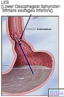
- Gastro-esophageal manometry allows to measure the time and strength of contractions and relaxations of the muscular valves at the level of the upper esophageal (SES) and lower (SEI) esophageal sphincter. More precisely, the test allows to highlight the association with the alterations of the motility or with the presence of a muscular hypertone, which determines an "increased resistance to movements:
- at the level of the upper esophageal sphincter, for Zenker's diverticulum;
- at the level of the lower esophageal sphincter, for epiphrenic diverticula.
- In case of evident symptoms such as the manifestation of dysphagia and odynophagia (painful sensation during swallowing), an EsophagusGastroDuodenoscopy (EGDS) is indicated, an endoscopic examination of the upper digestive tract, which allows to exclude structural pathological conditions associated with diverticula of the esophagus. , such as strictures or neoplasms.
Treatment
Typically, asymptomatic or minimally symptomatic diverticula do not need treatment.
In many patients with esophageal diverticula, dysphagia is related to the alteration of basic motility, so therapy must be directed to treat this disorder. For example, treatment of intramural esophageal diverticulum is directed at the underlying stricture or dysmotility.
Only in certain cases, in which the esophageal diverticulum reaches considerable size, or if it is associated with disabling symptoms, it is possible to evaluate the possibility of resorting to surgical removal (resection). The indications for the surgical treatment of esophageal diverticula are well represented by three characters: symptomatic, voluminous, disabling.
Therapeutic options may also include:
- Injection of botulinum toxin in the lower esophageal sphincter (with transient effect, from 1 to 3 months).
- Heller's esophageal myotomy (surgical resection of the bundles of smooth muscle tissue surrounding the esophagus).
Some surgical approaches are given below:

Zenker's diverticulum before and after esophagus-diverticulostomy - Source: http://stanfordhospital.org/
Surgery allows the stenosis to be resolved definitively, with clinical and radiological remission of the disease. In recent years, non-invasive techniques have been perfected which guarantee good results and modest post-operative pain.
Diet and Lifestyle
- Do not lie down (or bend over) immediately after main meals
- Sleeping with two pillows, to facilitate the emptying of the esophagus and limit the stasis of food
- Avoid large meals
- Avoid the intake of coffee, mint, chocolate, fatty foods and alcohol
- Reduce acidic foods that can irritate the walls of the esophagus: juices, citrus juices, tomato and pepper.

