See also: dental plaque
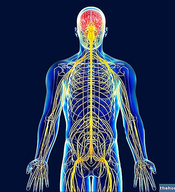
The neuromuscular plate allows the transmission of the nerve impulse between a termination of the motor nerve and the muscle. In response to this stimulus, muscle contraction occurs.
The final endings of the nerve fiber constitute the so-called presynaptic terminal. Their relationship with the external surface of the corresponding fiber (sarcolemma), called the postsynaptic surface, is not direct, but mediated by a space, called the synaptic space.

In order for the impulse to pass this space, the release of a neurotransmitter, specifically acetylcholine, by the presynaptic terminal is necessary; its task is to cross the synaptic space and deliver the "contractile message" to the muscle fiber.
The chemical synapse between nerve and muscle is called the NEUROMUSCULAR JUNCTION
After being poured into the synaptic space, acetylcholine is captured by specific receptors placed on the postsynaptic surface. The interaction between acetylcholine and receptor causes an increase in the permeability of the sarcolemma to sodium and potassium ions, resulting in a partial depolarization of the membrane postsynaptic. If this depolarization is large enough to exceed a certain threshold, the so-called action potential is triggered.
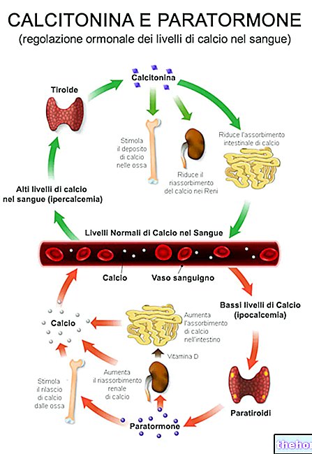
The action potential, thus generated, propagates inside the cell and the transverse tubules, thanks to the opening of the voltage-dependent Na + channels. The activation of receptors present in the membrane of these T tubules opens specific channels for the release of Calcium, located in the terminal cisterns of the sarcoplasmic reticulum.
The calcium released from the cisterns then diffuses into the cytosol, reaching concentrations 100 times higher than in the resting condition and initiating muscle contraction.The appearance of calcium near the Tn-C subunit of troponin causes the release of the active site on the actin and the consequent formation of actomyosin bridges.
Once the stimulus that gave rise to the contraction has ceased, relaxation occurs through an active ATP dependent process, which has the purpose of bringing calcium ions back into the sarcoplasmic reticulum, thanks to the action of a Ca2 + ATPase pump.
When the cytoplasmic concentration of free Ca2 + falls, the ion detaches from troponin, restoring the inhibitory effect of the troponin-tropomyosin system.
With reference to the action potential, it should be remembered that:
once generated, it determines the SYNCHRONOUS and MAXIMUM contraction of all the cells innervated by that motor neuron (it obeys the law of all or nothing).
Force regulation occurs through two main mechanisms:
1) increase in the number of motor units recruited;
2) variation of the discharge frequency of the motor neuron (repeated and close stimuli increase the intensity of the contraction and vice versa).
In regulating the force of contraction, the smallest motor units (red and slow fibers) are recruited first and then the larger ones (white and fast fibers).
summing up
1) An action potential travels along the axon of an alpha motor neuron to its terminations on a number of muscle fibers.
2) At the level of each termination, the nerve fiber secretes acetylcholine, which depolarizes the membrane of the muscle fiber, triggering the action potential
2) The propagation of the action potential induces the release of calcium at the level of the sarcoplasmic reticulum
3) Calcium binds to troponin C, eliminating the inhibitory effect on muscle contraction of the troponin-tropomyosin system
3) The muscle contracts, thanks to the hydrolysis of ATP by the myosinic heads and the subsequent traction on the thin actin filaments
4) Once the nervous stimulus ceases, calcium is reabsorbed by the tubular system and this, by activating the troponin-tropomyosin switch, switches off any further actomyosin interaction in the bud.
In addition to the afferent motor fibers, the muscle is also innervated by efferent sensory fibers. Sensory fibers include those of the neuromuscular spindles (sensitive to length) and those of the Golgi tendon organ (sensitive to tension), as well as a variety of free nerve endings, some of which are specific to pain perception.
Other articles on "Neuromuscular plaque"
- muscle innervation
- muscles of the human body
- Skeletal muscle
- Muscles classification
- Muscles with parallel bundles and pinnate muscles
- Muscle anatomy and muscle fibers
- myofibrils and sarcomeres
- actin myosin
- muscle contraction








-nelle-carni-di-maiale.jpg)
