Edited by Dr. Stefano Casali
The nervous system is anatomically organized into:
- Central Nervous System (CNS) includes the brain and spinal cord,
- Peripheral Nervous System (PNS) comprises the cranial nerves arising from the brain and the spinal nerves emerging from the spinal cord with the ganglia.
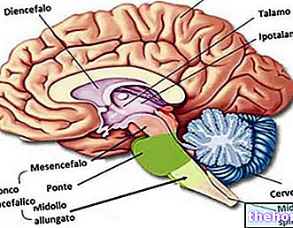
In "adult humans, the brain weighs an average of 1.3 to 1.4 kg. The brain contains about 100 billion * nerve cells (neurons) and trillions ** of" support cells ", called glia.
The spinal cord is about 43 cm long in the adult woman and 45 cm in the adult man and weighs about 35-40 g. The spine, the series of bones (in the back) that houses the spinal cord, is about 70 cm long. so that the spinal cord is much shorter than the spinal column.
(*) One billion equals 1,000,000,000,000, or 1012
(**) One trillion is equivalent to 1,000,000,000,000,000,000 or 1018
Central nervous system
The Central Nervous System is divided into two main parts:
- Brain;
- Spinal cord.
The brain is made up of gray matter and white matter. Its interior is mainly formed by a white matter, externally enveloped by a layer of gray matter, the cerebral cortex.
- The white matter is made up of myelinated fibers, oligodendrocytes, fibrous astrocytes and microglia cells. The white color is given by myelin;
- The gray matter contains the soma (cell body), unmyelinated and myelinated fibers, protoplasmic astrocytes, oligodendrocytes and microglia cells.
In the transverse sections of the spinal cord, the white matter is localized on the outside and the gray matter on the inside, where it assumes an H-shape. ependymal cells. The neural tube is a structure present in the embryos of the chordates, from which the central nervous system originates. Cylindrical in shape and equipped with a central cavity, the neural tube derives from a thickened region of the ectoderm, the neural plate, through a process called neurulation.
The gray matter forms the anterior horns of the containing H motor neurons from which the ventral roots of the spinal nerves originate. The dorsal horns of H are also receiving gray matter sensitive fibers from the neurons of the spinal ganglia.
- The anterior horn is made up of neurons responsible for motor functions (α motor neurons and γ motor neurons);
- while the posterior horn is given by neurons used for the sensory function, above all tactile and painful.
The CNS is protected by the skull and spine and also by connective tissue membranes called meninges. From the most external the meninges are:
- Hard mother;
- Arachnoid;
- Pious mother.
Peripheral Nervous System
The Peripheral Nervous System is divided into two main parts:
- Somatic Nervous System responsible for voluntary responses;
- Autonomous Nervous System, or Vegetative, responsible for involuntary responses.
The Somatic Nervous System is made up of peripheral nerve fibers that send sensitive information to the Central Nervous System and motor nerve fibers that lead to the skeletal muscles.
The Autonomous Nervous System is divided into two parts with antagonistic action:
- Sympathetic (thoracic - lumbar);
- Parasympathetic (craniosacral).
The Autonomous Nervous System controls the smooth muscles of the viscera and glands.
SYMPATHIC NERVOUS SYSTEM
The Sympathetic arises in the spinal cord. It stimulates the heart, dilates the bronchi, contracts the arteries and inhibits the digestive system, prepares the body for physical activity. Here, the cell bodies of the first neuron (the preganglionic neuron) are located in the thoracic and lumbar tracts. The axons originating from these neurons lead to a chain of ganglia located on either side of the spinal column (the lateral-vertebral ganglionic chain). In the ganglionic chain, most neurons synapse with another neuron (the post-ganglionic neuron). The post-ganglionic neuron then projects to the "target": a muscle (smooth or cardiac) or a gland.
In the sympathetic system, the preganglionic fibers are short, while the postganglionic fibers are long.
PARASYMPATHIC NERVOUS SYSTEM
It is called the Craniosacral Autonomous System since it refers to the viscero-motor nuclei of the encephalic nerves and to the viscera-effector sacral columns. The Parasympathetic is a system that predisposes to nutrition, digestion, sleep and rest. The centers of the Parasympathetic are found in the brain stem and in the sacral part of the spinal cord. In the brainstem there are the nuclei for the innervation of the salivary, nasal, lacrimal glands and of all the organs up to the left flexure of the colon which represents the border point between the middle intestine and the caudal intestine. In this system the preganglionic branches are long and reach the ganglia just outside or inside the organ to be innervated (for this reason the postganglionic fibers are very short). In the heart, the Parasympathetic has the task of decreasing the heartbeats, the pressure, and causing a vasoconstriction of the arteries of the heart (the coronaries). A coronary constriction results in a decreased blood supply to the heart. In the digestive tract, the Vagus represents the Parasympathetic and acts by causing peristalsis and, at the gastric level, the secretion of HCl.
ENTERIC NERVOUS SYSTEM
The Enteric Nervous System is an intrigue of nerve fibers that innervates the viscera (gastrointestinal tract, pancreas, gall bladder). In the various organs this acts through the plexuses (myenteric plexus and submucosal plexus).
Other articles on "Nervous System"
- neurons, nerves and blood-brain barrier
- nerve cells and synapses

.jpg)


