Generality
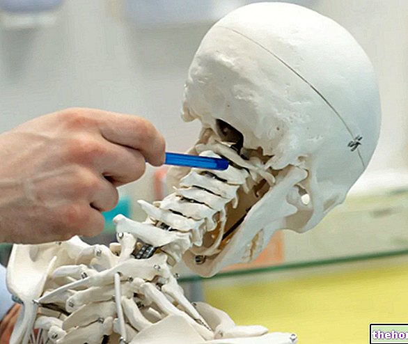
The brainstem is the most primitive region of the brain; it represents a sorting center for nerve signals, as the fibers that cross it travel to the spinal cord and the rest of the brain.
Furthermore, almost all the cranial nerves arise in the brain stem, which establish contacts with the muscles and organs of the head, neck, thorax and abdomen.

Figure: the brain and its main anatomical areas.
This article will address some of the main anatomical aspects of the brain stem, what its functions are and where the cranial nerves reside.
To understand the brain stem
To better understand how the brainstem is made, we need some premises.
THE ENCEPHALUS
Together with the spinal cord, the brain makes up the central nervous system (CNS). It is a very complex structure, as it is made up of several regions, each with a specific function.
In the "adult man," the brain weighs up to 1.4 kg (about 2% of the total body weight) and can contain 100 billion neurons (one billion corresponds to 1012). Therefore, the connections that it can establish are many and unimaginable.
There are four main regions of the brain: telencephalon, diencephalon, cerebellum and brainstem. Each of them has a specific anatomy, with compartments specialized in different functions.
REGION
FUNCTION
Cerebral cortex
Perception; movement and coordination of the voluntary muscles
Thalamus
Passage station for motor and sensory information
Instinctive behaviors; secretion of various hormones
Mid-brain
Eye movement; coordination of auditory and visual reflexes
NEURONS, NERVOUS FIBERS AND NERVES
Neurons are the cells that make up nerve tissue.
Thanks to their extensions (which make up the so-called nerve fibers), starting from the CNS they reach every region of the body.
The enormous communication network that neurons create allows them to perform their main function in the best possible way, which is to transmit signals of a nervous nature. Depending on the direction in which they perform this task, neurons have different roles and are distinguished in:
- Efferent or motor neurons (or motor neurons), if the signal travels from the CNS to the tissues (periphery)
- Afferent or sensory / sensory neurons, if the signal travels from the tissues (periphery) to the CNS
A bundle of neurons (or rather axons) can make up a nerve, which can be:
- Efferent nerve, i.e. consisting only of efferent neurons
- Afferent nerve, i.e. formed only by afferent neurons
- Mixed nerve, that is, composed of efferent and afferent neurons
CLARIFICATION ON THE TERMINOLOGY
In this and other texts relating to the brain and the central nervous system, it will often be possible to read opposing terms such as "ventral position - dorsal position", "rostral position - caudal position". What is their meaning? these concepts should be clarified.
If an anatomical element is located in a ventral position with respect to another, it means that it is in front of the latter and looks towards the belly of the human model taken as an example.
Conversely, if an anatomical component is placed in a dorsal position with respect to another, it means that it is behind the latter and is projected towards the back (or back) of the exemplary human model.
The rostral position is the upper side, oriented towards the head; while, the caudal position is the lower side.
Brain stem: general characteristics
The brainstem, or brain stem, is the oldest and most primitive region of the brain. Located under the diencephalon, it represents the nervous structure that connects the telencephalon with the spinal cord. Furthermore, although the so-called IV ventricle separates them. cerebral (a cavity), also establishes relations with the cerebellum, which is located in the dorsal position.
Seat of origin of almost all the cranial nerves, three regions can be recognized in the brain stem:
- the midbrain,
- the bridge (or bridge of Varolio)
- the medulla oblongata (or bulb).
INTERNAL ANATOMY AND MAIN FUNCTIONS

Figure: position and extent of the reticular formation. The cerebellum and IV ventricle sites can also be noted. From the site: drvannetiello.wordpress.com
In some respects, the internal anatomy of the brainstem resembles that of the spinal cord, so much so that it may appear to be a continuation of this.
The entire structure of the brain stem is crossed by neurons and by bundles of neurons with different functions. There are, in fact, bundles of motor neurons (or descending bundles), bundles of sensory neurons (or ascending bundles) and neurons that play a role of The latter make up the so-called reticular substance or reticular formation, which, located in the central region along the entire structure, is responsible for regulating certain activities controlled by the spinal cord, the cerebral cortex and the brainstem itself. The following processes depend on the action of the reticular formation:
- Sleep-wake cycles
- State of consciousness
- Muscle tone control
- Stretch reflexes
- Coordination of breathing
- Pain modulation
- Blood pressure regulation
THE CRANIAL NERVES AND THEIR FUNCTIONS
In the human brain there are twelve pairs of cranial nerves, identified with the Roman numerals from I to XII. Except the I and II pair (which arise, respectively, from the telencephalon and diencephalon), the remaining ten pairs originate in the brainstem.
Depending on the neurons that constitute them, the cranial nerves can be motor, sensory (or sensory) and mixed. They establish contacts with the muscles, glands and sense organs of the head and neck; the exception is the X pair, the vagus nerve, which unlike the others makes contact with various thoracic and abdominal organs.
Nerve
First name
Guy
Function
Site
THE
Olfactory
Sensory
Olfactory information (smell)
Telencephalon
II
Optical
Sensory
Visual information
Diencephalon
III
Oculomotor
Motor
Eye movements, pupillary constriction or dilation, accommodation of the lens
Mid-brain
IV
Trochlear
Motor
Eye movements
V.
Trigeminal
Mixed
Sensory information from the face; motor signals for chewing
Varolio bridge (or bridge)
YOU
Abducting
Motor
Eye movements
VII
Facial
Mixed
Gustatory sensitivity; efferent signals for the salivary and lacrimal glands; movements of the facial muscles
VIII
Vestibulocochlear
Sensory
Hearing and balance
IX
Glossopharyngeal
Mixed
Sensitivity of the oral cavity, baro- and chemoreceptors of blood vessels; efferents for swallowing and for the secretion of the parotid salivary gland
Medulla oblongata (or bulb)
X
Vague
Mixed
Afferents and efferents for many internal organs, muscles and glands
XI
Accessory
Motor
Muscles of the oral cavity, some muscles of the neck and shoulder
XII
Hypoglossal
Motor
Muscles of the tongue
Mid-brain
In close contact with the diencephalon (on the rostral side) and resting on the Varolio bridge, the midbrain is the smallest region of the brain stem. It contains the roots of the III and IV pair of cranial nerves, or, respectively, the oculomotor and the trochlear. It contains the centers responsible for visual reflexes (pupillary reflex, blinking) and auditory (adjustment of hearing at "intensity of sounds).
The most important anatomical elements of the midbrain are:
In the ventral position
- The cerebral peduncles: two in number, one on the right and one on the left (therefore lateral), are bundles of nerve fibers coming from the cerebral cortex and directed towards the spinal cord. In the middle of the peduncles, the third pair of cranial nerves emerges, which arise, however, in a very close ventral area.
- The tegmentum: is the site of reticular formation and other nerve fibers, known as red nuclei. Furthermore, it is the starting point of the III pair of cranial nerves.
In the dorsal position
- The roof (or quadrigeminal lamina): it is formed by structures that serve to transmit the nerve signal, called collicoli (upper and lower). Below the inferior colliculi are the roots of the 4th pair of cranial nerves.
In rostral position
- There substantia nigra (or black substance): composed of pars compacta and from pars reticulata, belongs to the midbrain only from an anatomical point of view, as it is controlled by regions of the telencephalon called nuclei of the base. For this reason and for the complex function that the black substance plays, this is not the most suitable place to talk about it.
- L "Silvio's aqueduct: it is a canal, visible through a cross section of the brain stem, which connects the IV cerebral ventricle with the III cerebral ventricle.
Bridge (or bridge of Varolio)
Placed above the medulla oblongata (or bulb), under the midbrain and in front of the cerebellum, the bridge of Varolius (or more simply bridge) is a large half-ring arranged crosswise. On average, it measures 27mm in height and 38mm in width.
Thanks to the nerve fibers that cross it, the bridge is responsible for sorting the traveling information on the brain-cerebellum axis. It also contains the breathing and sleep centers.
The most characteristic anatomical structures are:
In the dorsal position
- The tegmentum: continuation of the tegmentum of the midbrain, in addition to containing the reticular formation, it is the seat of four pairs of cranial nerves (V, VI, VII, VIII).
- The cerebellar peduncles: correspond to the points where the nerve fibers of the pons, but not only, enter the cerebellum.
In the ventral position
- The pontine nuclei (or nuclei of the base of the pons or nuclei of the pontine gray): they are, in fact, groups of nerve fibers whose starting point is the cerebral cortex and the point of arrival can be: the spinal cord, the medulla oblongata, the pons itself and the cerebellum.
Medulla oblongata (or bulb)
Located just below the pons and in front of the cerebellum, the medulla oblongata (or bulb) is the last section of the brain stem, before the spinal cord. It is formed by bundles of nerve fibers that connect the medulla with the brain and resembles, in shape, to the central portion of an inverted cone Its length is about 30 mm, while its width varies from 22-25 mm, in the rostral area, to 10-12 mm, in the caudal area.
Since it is also the point of origin of the X pair of cranial nerves, the bulb controls various visceral functions of the chest and abdomen, related to: breathing, blood pressure, swallowing, coughing and vomiting.
The most representative anatomical components are:
In ventral position
- The posterior cords (or nuclei of the dorsal columns): are aggregates of nerve fibers, located in the lowest part (caudal position) of the inverted cone representing the medulla oblongata. Connected to the cerebellum through the cerebellar peduncles, the posterior cords are called tubercles (gracilis and cuneate), in the most rostral part, and fascicles (gracilis and cuneate), in the most caudal part. It is at the level of the tubercles, which are the roots of the XII and X pairs of cranial nerves.
In ventral position
- The two bulbar pyramids (right and left): they are two large clusters of nerve fibers, which come from the cerebral cortex and cross in the middle of their journey, or those on the left go to the right and vice versa. This intersection, called decussation, explains why the stimuli coming from the right side of the body are transmitted to the left side of the brain, which in turn sends stimuli to the right side of the body (and vice versa for the opposite side). of the pyramids, the extensions of the cranial nerves of the XII pair emerge.In the passage from medulla oblongata to spinal cord, the pyramids disappear.
- The right and left olive complex: they are two substantial masses of nerve fibers that come from the cortex, the spinal cord and the midbrain and head towards the cerebellum. Behind the olive complexes, the branches of the cranial nerves of the XI, X and IX pair protrude.

Figure: ventral view of the brain stem. In blue, the cranial nerves have been highlighted; in green, the midbrain and its cerebral peduncles (the tegment is visible only through a cross section of the brain stem, as it is contained within); in black, the bridge and its Pontine nuclei; in red, the medulla oblongata, its bulbar pyramids, its olive complex and the decussation of the pyramids. From the site: mussejereissati.com

Figure: dorsal view of the brain stem. In blue, the cerebellum (of which only a part can be seen, for reasons of convenience) and the cavity in which the IV cerebral ventricle resides are highlighted; in green, the midbrain and its collicles; in black, the bridge, the site where the pontine cranial nerves are born and the position of the cerebellar peduncles; finally, in red, the medulla oblongata and its posterior cords, separated by the so-called posterior sulcus. From the site: med.ufro.cl


