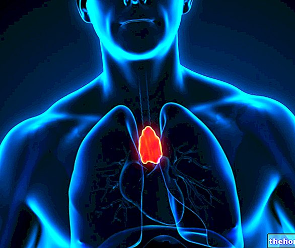
An uneven, asymmetrical and flat organ, the thymus is located in the anterior superior mediastinum, lying on the pericardium, behind the breastbone and anterior to the large blood vessels that branch off from the heart.
The thymus is characterized by an important growth and by an intense activity until puberty, after which, due to the effect of the sex hormones, it becomes smaller and less active.
The thymus is the organ responsible for maturing the T lymphocytes produced in the bone marrow and transferred to the same thymus during fetal life.
The thymus is a particular organ, which in the course of life changes its size and its composition, until it becomes, in adulthood, a small and mainly adipose structure.
, and below the sternohyoid and sternothyroid muscles.
In the period of maximum development (puberty), the thymus extends from the lower pole of the thyroid to the fourth pair of costal cartilages.
Microscopic Anatomy: Histology of the Thymus

The thymus has a layer of superficial connective tissue, high in collagen and reticular fibers, called a capsule.
Therefore, under the capsule, in each lobule, it is possible to recognize two distinct cellular components, one more external and one more internal:
- The outermost cellular component is the so-called cortical zone (or cortex).
Dark in color under the microscope, the cortical zone of the thymus contains large quantities of thymocytes, reticular epithelial cells and macrophages. - The innermost cellular component is the so-called medullary zone.
Light in color under the microscope, the medullary area of the thymus contains a small amount of thymocytes and, in contrast, an "abundance of reticular epithelial cells, some of which are organized in structures called Hassall's corpuscles.
What are thymocytes?
Thymocytes are the cells of the thymus responsible for giving rise to T lymphocytes; they are, therefore, the precursors of T lymphocytes.
As will be seen later, thymocytes are formed in the bone marrow and are transferred to the thymus, for subsequent maturation into T lymphocytes, in the most advanced stages of the embryonic formation of the thymus itself.
What are epithelial reticular cells?
Constituting the so-called thymic epithelium, reticular epithelial cells (or thymic epithelial cells) are the cellular elements that make up the parenchyma of the thymus (the parenchyma is the functional component of an organ).
The reticular epithelial cells contain granules, which appear to house thymic hormones.
Reticular epithelial cells play a key role in the maturation process of thymocytes to T lymphocytes.
What are the Hassall Corpuscles?
Hassall corpuscles are concentrically arranged formations of reticular epithelial cells filled with keratin filaments.
Their functional role is not yet known precisely.
Vascularization of the Thymus
The supply of oxygenated blood to the thymus belongs to the branches (or branches) of the internal thoracic artery, the inferior thyroid artery and, sometimes, the superior thyroid artery.
The internal thoracic artery is a direct derivation of the subclavian artery; the inferior thyroid artery derives from the thyrocervical trunk, which in turn emerges from the aforementioned subclavian artery; finally, the superior thyroid artery is a branch of the external carotid artery.
As for the venous blood leaving the thymus, this flows into the left brachiocephalic vein, the internal thoracic vein and the inferior thyroid vein; it should be noted, however, that, in some individuals, the venous blood leaving the thymus flows directly, through small veins, into the superior vena cava.
The left brachiocephalic, internal thoracic, and inferior thyroid veins all flow into the superior vena cava.
Lymphatic Circulation of the Thymus
The thymus has no afferent lymphatic vessels (ie which reach the thymus), while it has several efferent lymphatic vessels (ie which depart from the thymus).
The efferent vessels of the thymus are responsible for draining the lymph in the lymph nodes located near the thymus itself; such lymph nodes are:
- The mammary-parasternal lymph node;
- The tracheobronchial-hilar lymph node;
- The mediastinal-brachiocephalic lymph node.
Innervation of the Thymus
The innervation of the thymus is minimal.
To innervate the thymus are branches (or branches) of the vagus nerve, branches of the cervical segment of the so-called sympathetic chain and branches of the phrenic nerve (these are limited to the innervation of the portion called the capsule).
and the parathyroid.At the 8th week of gestation, the thymic epithelium moves up to assume the position of the thymus during life, ie at the level of the anterior superior mediastinum.
Once the anterior superior mediastinum is reached, the thymic epithelium starts the formation of lobules, which ends with the generation of the thymus proper.
The thymocytes, on the other hand, begin to make their appearance at a much more advanced gestational age (compared to the thymic epithelium); usually, the first thymocytes appear during the formation of thymic lobules.
To give rise to the thymocytes is a line of cells deriving from the bone marrow (pre-thymocytes), which, precisely for the transformation into thymocytes, is transferred to the level of the future thymus.
Scientific studies have shown that the origin of thymocytes is crucial for the completion and further development of the thymic epithelium.
Did you know that ...
Iodine is crucial for the development and activity of the thymus.
Evolution of the Thymus over the course of life
From birth to puberty, the thymus grows in size, reaching to weigh, at the peak of its size, even 40-50 grams (at birth it weighs around 12 grams).
The increase in size of the thymus coincides with its greater activity.
With puberty, therefore, the thymus begins a process of involution (thymic involution), which decrees a drastic reduction in size and a change in composition such that the functional tissue takes over the adipose tissue.
At the end of thymic involution, the thymus becomes a small and much less active organ than in the years before puberty.
What causes thymic involution?
The involution of the thymus is due to the increase in the levels of circulating sex hormones, which typically occurs with the onset of puberty.
This process, however, can also recognize non-physiological causes; among all, we point out AIDS, that is the infectious disease caused by the human immunodeficiency virus (HIV).
Did you know that ...
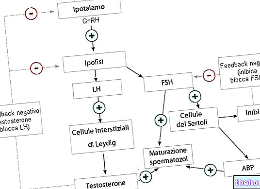
Chemical castration could reverse the process of thymic involution and restore the activity of the thymus. Moreover, chemical castration inhibits the activity of the gonads, which are the endocrine organs responsible for the production of sex hormones.
which contribute to the so-called cell-mediated immunity.Cell-mediated immunity - which, in addition to T lymphocytes, also includes macrophages, cells natural killer and cytokine-secreting cells - is part of adaptive immunity and serves primarily to:
- Remove virus infected cells;
- Eliminate fungi, protozoa, tumor cells and intracellular bacteria;
- Destroy surviving microbes from phagocyte activity (macrophages, neutrophils, monocytes, dendritic cells and mast cells).
Did you know that ...
Cell-mediated immunity is the component of the immune system that intervenes in the rejection process of a transplanted organ.
T lymphocyte maturation: the details
The maturation of T lymphocytes operated by the thymus can be divided into two phases: a first phase, called positive selection, and a second phase, called negative selection.
Positive Selection
During the positive selection, we witness:
- The creation of peptide receptors whose destiny is to attach themselves to the surface of future T lymphocytes and act as an antigen recognition structure (an antigen is any substance foreign to the organism, which can threaten its state of health).
- The elimination of potential T lymphocytes presenting, on the surface, non-functional peptide receptors; it may happen, in fact, that the process of creation of the aforementioned receptors makes mistakes and that these errors result in potential T lymphocytes unable to recognize the antigens (non-functional ).
The selection between functional and non-functional T lymphocytes sees as protagonist a set of molecules known as major histocompatibility complex (MHC); by actually replicating the known antigens that could threaten the organism, MHC is able to test which T lymphocytes possess the ability to recognize the antigen and which, on the contrary, do not.
The recognition capacity test is based on a "binding affinity between the MHC itself and the potential T lymphocytes: if the T lymphocytes bind to MHC they pass the control and progress in maturation; if they do not bind, instead, they do not pass the control and undergo apoptosis (programmed cell death). - The targeting of T lymphocytes that have passed control to the CD8 (cytotoxic T lymphocytes) or CD4 (helper T lymphocytes) lymphocytes.
Positive selection takes place at the level of the cortical area of the thymus: it is, in fact, the reticular epithelial cells present here that carry out the aforementioned processes.
Negative Selection
Positive selection ensures that potential T lymphocytes are able to recognize antigens, but not that they are also reactive to the organism's own molecules (autoantigens).
The reticular epithelial cells of the medullary area of the thymus are responsible for identifying and subsequently eliminating the T lymphocytes that recognize autoantigens; this fundamental process for the well-being of the organism is negative selection.
In the absence of an appropriate negative selection, the T lymphocytes capable of reacting against the autoantigens would survive and damage organs and tissues of the organism to which they belong.
The effects just described are called self-reactivity; self-reactivity is the pathophysiological mechanism underlying autoimmune diseases.
Molecules involved in the maturation of T Lymphocytes: Thymic Hormones
Some hormones secreted by the thymus itself also contribute to the maturation process of T lymphocytes; among these hormones, thymosin, thymopoietin and thymulin are reported.
It is by virtue of its ability to produce the aforementioned hormones that the thymus is part of the endocrine glands.
Cytotoxic T Lymphocytes and Helper T Lymphocytes
Given their importance and since they have been named, it is necessary to provide the reader with some more details about cytotoxic T lymphocytes and helper T lymphocytes:
- CD8 cytotoxic T lymphocytes: these are T lymphocytes capable of recognizing infected cells and destroying them in the first person.
- CD4 helper T lymphocytes: these are T lymphocytes that coordinate an immune response only on stimulation of other cells of the immune system (macrophages, B lymphocytes and dendritic cells); the response they trigger, moreover, consists in a release of cytokines, whose fate is to activate further other elements of the immune system (eg: leukocytes, memory B cells, etc.).
CD4 T helper lymphocytes are, therefore, modulators of the immune response.
What Happens After T Lymphocyte Maturation?
Once their maturation is complete, the lymphocytes leave the thymus and spread into the blood, lymph and secondary lymphoid organs (e.g. spleen, lymph nodes and tonsils).
Why does the physiological involution of the thymus not expose you to infections?
As described above, at some point in life (puberty), the thymus becomes smaller and almost completely loses its activity (thymic involution).
The physiological involution of the thymus, however, does not compromise the efficiency of the cell-mediated immunity implemented by the T lymphocytes and does not in any way determine a greater exposure to infections. Here are the reasons:
- Until puberty, the thymus is so active that it also produces T lymphocytes for future years of adult life;
- The activity that the thymus retains in adulthood is minimal, but still sufficient to keep intact the T lymphocyte patrimony produced in the first years of life.
What has just been described does not apply, of course, in the case of an "early thymic involution: when the thymus regresses earlier than expected, there is not" the time necessary to build a patrimony of T lymphocytes that can also be spent in future years, therefore the individual concerned will be more sensitive to infections.
DiGeorge's, myasthenia gravis and thymic cysts.Thymoma
Thymoma is the name of any tumor that arises from the uncontrolled proliferation of one of the epithelial cells of the thymus.
Generally, thymoma is a malignant tumor and remains so; although rarely, however, it can transform into a malignant form and become an invasive and very dangerous carcinoma.
Associated in 20% of patients with myasthenia gravis, thymoma mostly affects adults over 40 years of age and of Asian ethnicity.
Due to the mass effect of the tumor, the typical symptoms and signs of thymoma consist of: compression of the vena cava, dysphagia, cough and chest pain.
For the diagnosis of thymoma, imaging tests such as CT scans, MRIs, and X-rays are essential.
Among the treatments that can be adopted in case of thymoma, there are surgery, chemotherapy and radiotherapy.
DiGeorge syndrome
DiGeorge syndrome is a genetic disorder characterized by the absence (deletion) of a portion of chromosome 22.
Due to the absence of a stretch of chromosome 22, DiGeorge syndrome is associated with numerous congenital malformations, including aplasia of the thymus.
Thymus aplasia consists in the absence of the thymus and involves a form of primary immunosuppression, which clearly exposes the patient to repeated infections.
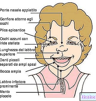
DiGeorge syndrome also causes congenital heart defects, facial abnormalities, cleft palate, and hypoparathyroidism.
Myasthenia Gravis
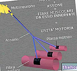
Myasthenia gravis is a chronic disease characterized by fatigue and weakness of some muscles of the human body.
Myasthenia gravis is an autoimmune disease; it is due, in fact, to the presence of some autoantibodies that block the post-synaptic receptors of the neuromuscular junction and thus inhibit the excitatory effects of acetylcholine.
At least for some patients, the thymus seems to play a role in the etiology of myasthenia gravis: in an important percentage of cases, in fact, there is an abnormal enlargement of the thymus (hyperplasia) and / or the appearance of a thymoma.

