Generality
The skeleton is the internal scaffolding of the human body. In its constitution, mainly the bones participate and, secondly, the cartilages and joints.

The man has a slightly different skeleton from the woman: the differences are subtle, however an expert eye (eg: a doctor) is able to grasp them and understand the sex of an individual from the sole observation of the skeletal framework (when, clearly, no other information is available).
The skeleton covers various functions, including: support of the human body, protection of underlying organs and soft tissues, assistance with balance and movement, production of blood cells, release of the osteocalcin hormone and storage department for mineral salts such as calcium and iron.
The skeleton can be the victim of injuries (eg: bone fractures or joint sprains) and pathologies, such as osteoporosis or arthritis.
What is the skeleton?
The skeleton is the internal scaffolding of the human body, in which the bones (main component), cartilage tissues and joints participate.
Anatomy
The skeleton of an adult human being constitutes 30-40% of the total mass of the body (body mass) and includes as many as 206 bones, different in shape and function, and present in equal (eg: the two femurs) or uneven (eg. : hyoid bone).
ANATOMICAL DIVISIONS: AXLE AND APPENDICULAR SKELETON
According to the classical anatomical view, the skeleton of the human being can be divided into: axial skeleton and appendicular skeleton.
The axial skeleton is the set of bones that make up the skull, the vertebral column and the rib cage, plus the hyoid bone and the three ossicles of each ear (hammer, anvil and stirrup). In all, it includes 80 bone elements:
- The 22 bones of the skull;
- The 26 bones of the vertebral column, as long as the bones of the sacral tract (or sacral vertebrae) are considered as one and constituting the so-called sacrum (otherwise, the bones of the vertebral column would be 33-34);
- The 25 bones of the rib cage (12 pairs of ribs plus the breastbone).
- The aforementioned hyoid bone and 3 ossicles of each ear;
The appendicular skeleton, on the other hand, represents the set of bones that form the shoulder girdle (or shoulder girdle), the upper limbs, the pelvis and the lower limbs. Overall, it includes 126 bone elements:
- The 4 bones of the shoulder girdle, which are the 2 shoulder blades and the 2 collarbones;
- The 3 bones of each upper limb hand excluded, which are humerus, radius and ulna;
- The 27 bones of each hand, which are the carpal bones, metacarpals and phalanges of the fingers. The two hands, therefore, contain the beauty of 54 bones;
- The 2 bones of the pelvis, which are the iliac bones;
- The 4 bones of each lower limb excluding foot, which are the femur, the patella, the tibia and the fibula;
- The 26 bones of each foot, which are the tarsal bones, metatarsals and phalanges of the toes. The two feet, therefore, contribute to the total number of bones of the skeleton with as many as 52 elements.
COMPOSITION OF THE BONES
The bones of the skeleton are the result of a cellular component and a non-living component, called the bone matrix.
- The cellular component of skeletal bones comprises three types of cells, which are: osteoblasts, osteoclasts and osteocytes. The contribution of the cells just mentioned, to the total mass of the skeleton, is small; this does not, however, mean that they have a "fundamental importance for the health of the bones and the skeleton in general.
- Turning to the bone matrix, this is half water and half collagen mixed with calcium phosphate (83-85%), calcium carbonate (9-11%), magnesium phosphate (1-2%) and calcium fluoride (0.7-3%). It should be noted that, often, calcium phosphate, calcium carbonate and calcium fluoride, present in the bones, are known with a more general term, corresponding to hydroxyapatite.
To learn more about the cellular component of skeletal bones, readers can consult the article here.
TYPES OF SKELETON BONES
Based on shape and size, anatomists distinguish the bones of the human skeleton into at least 6 different types, which are:
- The typology of the long bones. All bones in which length prevails over width and thickness belong to this category. Long bones are distinguished by a narrow central part, called the diaphysis or body, and by two bulky ends, called the epiphysis.
Inside the long bones, to be precise inside the diaphysis, resides the bone marrow, whose function will be considered in the chapter dedicated to the functions of the skeleton.
The bone tissue that makes up long bones is generally very compact.
Typical examples of long bones are: the humerus, the ulna, the radius, the femur, the tibia, the fibula and the clavicle. - The type of short (or short) bones. Bones in which length and diameter are equivalent belong to this category.
The short (or short) bones have a particular composition: spongy bone tissue, internally, and compact bone tissue, externally.
Typical examples of short (or short) bones are: the wrist bones, the calcaneus and the vertebrae. - The typology of flat bones. All bones of limited thickness and laminar appearance fall into this category.
Despite their thinness, flat bones consist of two layers of bone tissue: an inner layer, which includes spongy bone and bone marrow, and an outer layer, which includes compact bone.
Classic examples of flat bones are: the bones of the skull, pelvis and sternum and the shoulder blades. - The typology of irregular bones. Irregularly shaped bones belong to this category and are difficult to describe.
Two examples of irregular bones are the ethmoid and the sphenoid, two bones of the splanchnocranium. - The typology of the sesamoid bones. All small, rounded and flattened bones fall into this category.
The sesamoid bones are important for the relationship they establish with the tendons.
The most classic example of sesamoid bone is the patella of the knee. - The typology of the bones wormian or sutural. Flat and indefinitely shaped bones, found between the sutures of the skull bones, belong to this category.
CARTILAGE TISSUES
Cartilage tissues, better known as cartilage or cartilage (in the singular), are supporting connective tissues, endowed with extreme flexibility and resistance.
Without blood vessels, cartilages are tissues that result from the union of particular cells, called chondrocytes.
In the human skeleton, cartilage tissues can have different peculiarities, depending on the functions they must perform. To understand what has just been said, the reader should think of the cartilage of the auricles and the cartilage of the menisci: although belonging to the same category of tissue and even resulting from the "union of chondrocytes, these two examples of cartilage differ considerably in consistency and specific properties.
The skeleton of the human being includes three types of cartilage:
- The hyaline cartilage;
- The elastic cartilage;
- Fibrous cartilage.
It is not present in the joints.
It is richly present at the joint level.
ARTICULATIONS
Joints are anatomical structures, sometimes complex, which put two or more bones into mutual contact. In the human skeleton, they are 360 and fulfill functions of support, mobility and protection.
According to the most common anatomical view, there are three main categories of joints:
- The fibrous joints (or synarthrosis). They generally lack mobility and the constituent bones are held together by fibrous tissue. Typical examples of synarthrosis are the joints present between the bones of the skull.
- The cartilage joints (or amphiarthrosis). They have little mobility and the constituent bones are joined by cartilage. Classic examples of amphiarthrosis are the joints that connect the vertebrae of the spine.
- The synovial joints (or diarthrosis). They have great mobility and include various components, including: the joint surfaces and the cartilage that covers them, the joint capsule, the synovial membrane, the synovial bags and a series of ligaments and tendons.
Typical examples of diarthrosis are the shoulder, knee, hip and ankle joints.
DIFFERENCES BETWEEN THE TWO SEXES
The skeleton of the man has some differences compared to the skeleton of the woman.
These differences are subtle (only an expert eye is able to grasp them) and concern:
- The skull. Between the male skull and the female skull there is a gross difference at the level of: midline nuchal, mastoid processes, supraorbital margins, superciliary arches and chin.
- The long bones and the musculature that concerns them. The long bones of men are wider than the long bones of women. Furthermore, the areas of insertion of the muscles, on the long bones, are much wider and more resistant in men than in women, demonstrating the greater muscular strength of the male. male compared to female.
- There pelvis. The female pelvis differs from the male pelvis in shape and size. In fact, it is wider and more spacious, to allow the growth of the fetus, during a possible pregnancy, and favor the exit of the same fetus at the time of delivery. Therefore, the differences in the pelvic area between the two sexes are linked to reproduction.
In the presence of a skeletal remnant whose gender belonging is unknown (is it a man or a woman?), The observation of the pelvis represents one of the most accurate and reliable methods of investigation to establish sex. - There general strength of the skeletal scaffolding. Female skeletal elements have a tendency to be less robust and smaller than equivalent male skeletal elements.
The skeletal differences between men and women are an example of sexual dimorphism.
By sexual dimorphism, s "means the morphological difference between individuals belonging to the same species, but of different sexes.
Maybe the readers don't know that ...
In the human skeleton, a long bone that makes it possible to establish, with a certain degree of confidence, the sex of an individual is the collarbone.
Compared to the female clavicle, the male clavicle is thicker, forms a more accentuated S, lacks symmetry (in the sense that the right clavicle is different from the left clavicle) and, finally, has areas of insertion for the wider muscles.
SKELETON IN BABIES
The skeleton of a newborn human being comprises about 300 bones, which is nearly a hundred more bones than the skeleton of an adult human.
This difference is due to the fact that, with growth, many distinct adjacent bones fuse together, forming a single bone.
Typical examples of bones that fuse, during growth, are the bones of the skull (process of fusion of the cranial sutures).
Development
Over the course of life, the human skeleton undergoes several changes.
As stated, it changes in the number of bones, as a result of fusion processes; it also changes in its composition, which passes from predominantly cartilaginous, during fetal life and in the first years of existence, to predominantly bone, in adult life; finally, it changes in size, due to an increase in length and diameter of the bones.
Functions
The skeleton fulfills a variety of functions, including:
- Support. The bony elements of the so-called axial skeleton are indispensable for maintaining an upright posture and for the correct discharge of weight from the upper body (head, trunk and upper limbs) to the lower part of the body (hips and lower limbs).
- Protection of organs and delicate soft tissues. This is the case of the cranium (or bones of the skull) towards the brain, the rib cage towards the organs located in the thorax (heart, lungs, aorta, etc.), the vertebrae towards the spinal cord and the bones of the pelvis towards the abdominal organs.
- Balance and movement, along with muscles and nerves. The bones of the appendicular skeleton mainly provide balance and movement.
- Production of blood cells (red blood cells, white blood cells and platelets). The process of producing blood cells belongs to the bone marrow, present inside the long bones, and is called hematopoiesis.
- Plastic. The skeleton of each individual gives a very precise shape to the body of the latter.
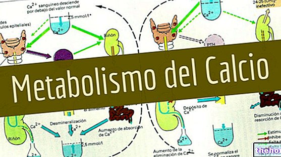
- Deposit of mineral salts. The bones of the skeleton are essential for the storage and metabolism of calcium, for the metabolism of iron and for the accumulation of iron in the form of ferritin.
This is not surprising, if you think back to the so-called bone matrix, rich in calcium phosphate, calcium carbonate, etc. - Release of the hormone osteocalcin. The main tasks of osteocalcin are: to increase insulin secretion, acting directly on the pancreas, and to increase insulin sensitivity, by acting on fat cells.
Clinic
The skeleton can be the victim of injuries and various pathologies.
Among the injuries of the skeleton, we report, first of all, bone fractures and, secondly, sprains / joint dislocations.
Among the pathologies of the skeleton, however, certainly deserve a mention: osteoporosis, osteopenia and arthritis.
BONE FRACTURES AND JOINT DISTORTIONS
Bone fractures and joint sprains / dislocations are injuries to the skeleton, which generally have a traumatic origin. The former concern the bones, while the latter concern the joints.
Typical symptoms of bone fractures and joint sprains / dislocations are: pain, limitation in movement (e.g. lameness, if lower limbs are involved), swelling and hematoma.
Treatment depends on the severity of the injury: minor injuries heal with rest, a cast (in the event of a fracture) and physiotherapy, while serious injuries require the intervention of the surgeon (in addition to rest, plaster cast and physiotherapy ).
OSTEOPOROSIS AND OSTEOPENIA
Osteoporosis is a common systemic disease of the skeleton, which causes a strong weakening of the bones. This weakening originates from the deterioration of the microarchitecture of the bone tissue and the consequent reduction of the bone mineral mass (eg reduction of calcium levels and / or iron, etc.) As a result of the aforementioned bone weakening, the bones of people with osteoporosis are more fragile and prone to fractures.
Osteopenia is a very similar condition to osteoporosis; to distinguish it from the latter are the lower degree of reduction in bone mineral density and the consequent lower risk of skeletal fractures. In other words, osteopenia is a mild form of osteoporosis.
Osteopenia and osteoporosis are two typical conditions of old age: in the female population, it is particularly common from the age of 65 onwards, in the male population, on the other hand, it is particularly common from the age of 70 onwards.
ARTHRITIS
The term arthritis refers to any inflammatory condition that affects one or more joints of the skeleton.
There are several types (or forms) of arthritis, each with its own unique causes and characteristics.
Among the most well-known and widespread types of arthritis, certainly deserve a mention: osteoarthritis (or arthrosis), rheumatoid arthritis, gouty arthritis (or gout) and ankylosing spondylitis.
The classic symptoms of arthritis are: pain, joint stiffness, joint swelling, redness and a sense of heat in the affected joint and, finally, reduced ability to move on the part of the joint involved.
Arthritis is a very common morbid condition of the skeleton which, in its various forms, can affect individuals of any age.