Generality
The coxa valga is the deformity of the hip in which the angle existing between the head-neck complex of the femur and the body of the femur measures at least 140 degrees, ie at least 5 degrees more than what represents normalcy.

Image taken from en.wikipedia.org
Example of valgus, the coxa valga can be the result of some neuromuscular diseases (eg: cerebral palsy), some skeletal dysplasias (eg: mucopolysaccharidosis) or hip trauma at a young age, such as to alter the normal growth process of the femur .
In coxa valga carriers, the presence of symptoms depends on the degree of the deformity: if the deformity is mild, the coxa valga is asymptomatic; on the contrary, if the deformity is severe, the coxa valga is responsible for various disorders (eg: lameness, hip pain, joint stiffness, etc.) and can give rise to serious complications (eg: osteonecrosis of the femoral head).
The diagnosis of coxa valga is generally based on: the physical examination, the medical history and a radiological examination referred to the hip.
In the presence of coxa valga, the use of a therapy and the type of therapy adopted depend on the presence or absence of symptoms.
Brief reminder of valgus
Valgus is the orthopedic term that indicates limb deformities in which, due to an abnormal relationship between two adjacent bones or between two contiguous portions of the same bone, the most distal of these two it presents an orientation lateral, that is, it tends to move away in an atypical way from the sagittal plane.
The presence of valgus can have various consequences on the joint in which the deviated skeletal element participates, consequences that can be of an anatomical nature and, in the most serious cases, also of a functional nature. Furthermore, a certain painful symptomatology may also depend on valgus.
- The femur is proximal to the tibia, which is proximal to the foot bones;
- In the femur, the end bordering the trunk is the proximal end.
It means "farther from the center of the body" or "farther from the point of" origin ".
Examples:
- The tibia is distal to the femur;
- In the femur, the end bordering the knee is the distal end.
Example:
- The first toe (big toe) is medial to the other toes.
Example:
- The second, third, fourth and fifth toes are all lateral to the big toe.
What is the coxa valga?
Coxa valga is the name of the hip deformity in which the characteristic angle existing between the head-neck complex of the femur and the body of the femur measures at least 140 degrees, that is, at least 5 degrees more than the maximum limit that defines the normal range. for the angle in question.
The angle existing between the head-neck complex of the femur and the body of the femur is normal when its gradation is between 120 and 135 degrees.
Why is it an example of valgus?
The coxa valga is an example of valgus, since, due to the greater gradation of the angle existing between the head-neck complex of the femur and the body of the femur, the latter has a tendency to assume a more lateral orientation than normal. , in order to compensate for the greater amplitude of the aforementioned angle.
The coxa valga can be mono- or bi-lateral
Coxa valga can be a problem limited to one hip only (coxa valga unilateral) or involving both (coxa valga bilateral).
As a rule, the triggering factor, i.e. the cause, affects the involvement of one or both joints.
Is it the opposite of coxa vara?
The coxa valga is the deformity of the hip opposite to the coxa vara. According to medical definitions, in fact, the coxa vara is the "anomaly of the hip" in which the angle existing between the proximal end of the femur and the body of the femur it measures less than 120 degrees, ie less than the minimum limit that establishes the normal range for the angle in question.
The coxa vara is an example of varus, the condition contrary to valgus.
Causes
Possible causes of coxa valga include:
- Neuromuscular diseases, such as cerebral palsy, poliomyelitis and spinal dysraphism;
- Some forms of skeletal dysplasia, including mucopolysaccharidosis and Turner syndrome;
- Trauma to the hip at a young age, such as to interfere with the correct growth process of the femur.
While the aforementioned neuromuscular diseases and the aforementioned forms of skeletal dysplasia are more often responsible for bilateral coxa valga, trauma to the hip such as to interfere with the normal growth process of the femur are more frequently associated with unilateral coxa valga.
Symptoms and complications
In people with coxa valga, the presence of symptoms and signs is strictly dependent on the severity of the joint deformity. In fact, when it is mild, the coxa valga tends to be asymptomatic; conversely, when severe, it is generally associated with several symptoms and signs, including:
- Pain in the hip (if the deformity is unilateral) or in both hips (if the deformity is bilateral);
- Loss of joint mobility on the part of the hips
- Joint stiffness felt in one or both hips, depending on whether the deformity is mono- or bi-lateral;
- Lameness
- Spasticity of the hip adductor muscles, due to their abnormal overdevelopment (especially when compared with the abductor and extensor muscles of the hip);
- Shortening of one or both lower limbs, depending on whether the deformity is, respectively, unilateral or bilateral (clearly, in the unilateral coxa valga, the lower limb that undergoes the shortening is the one carrying the deformity);
- Knee varus. The varus knee is the deformity of the lower limbs that reflects a misalignment of the femur and tibia, such that the two knees point outward, bones in the opposite direction to each other.
Complications
The most severe cases of coxa valga can degenerate into episodes of osteonecrosis of the femoral head, into events of dislocation or subluxation of the femoral head and into very painful pressure ulcers (or bedsores).
Curiosity: what is osteonecrosis?
Osteonecrosis is the death of bone tissue due to a lack or insufficient blood supply.
Also known as avascular necrosis, bone necrosis or bone infarction, osteonecrosis results in the appearance of tiny fractures in the affected bone tissue and, especially in severe cases, the so-called phenomenon of bone collapse.
Diagnosis
As a rule, a thorough physical examination, a thorough medical history, and instrumental examination such as radiography of the hip are essential for an accurate and safe diagnosis of coxa valga.
What is the purpose of the physical examination?
The physical examination consists in the medical observation of the symptoms and signs revealed by the patient.
Very often, it includes the execution, by the patient and on the doctor's recommendation, of specific maneuvers or gestures, which serve to bring to light certain characteristic signs of the present pathological condition.
What is the anamnesis for?
The anamnesis (or clinical history) is the critical study of the symptoms and all the facts of medical interest, reported by the patient or by the family members of the latter.
In the case of coxa valga, the anamnesis is essential to trace the causes of the deformity; it is in fact through the investigation of the clinical history that the doctor becomes aware of any past trauma to the hip, the presence of neuromuscular diseases, etc.
What can be any in-depth exams?
If the hip radiograph is not comprehensive and does not allow a definitive diagnosis, doctors may order more in-depth imaging tests, such as nuclear magnetic resonance or CT of the pelvis.
What is the appearance of the femur on radiological images?
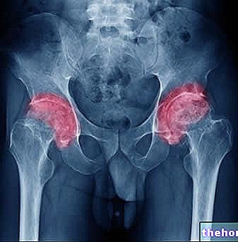
Radiological images of the hip of an individual with coxa valga show the head of the femur tending to align with the body of the femur. This anomaly is the normal consequence of the greater amplitude of the angle existing between the two femoral portions observed.
Therapy
For cases of asymptomatic coxa valga, no treatment is needed.
For mildly symptomatic cases of coxa valga, the canonically provided treatment is conservative and consists of physiotherapy.
Finally, for severely symptomatic cases of coxa valga, the only therapy currently effective is a surgical procedure known as femoral osteotomy with variating effect (or varizing femoral osteotomy).
Femoral osteotomy with variating effect
A rather delicate operation, the "femoral osteotomy with varizing effect involves the remodeling of the proximal portion of the femur, in order to reduce the present valgus (NB: deriving from varus, the opposite condition to valgus, the term" variating effect "refers precisely to the aforementioned purpose).
In the presence of a severely symptomatic coxa valga, the risk / benefit ratio of a delicate operation such as femoral osteotomy with a variating effect tends in favor of the latter. In other words, when the coxa valga is severe and very debilitating, it is better to operate and run the risks of surgical therapy, which do not leave free space for possible complications of the present deformity.
Prognosis
In the case of coxa valga, the prognosis depends on several factors, including:
- The degree of severity of the deformation. The more severe the deformation, the more difficult the treatment;
- The timeliness of the treatment. Failure to treat coxa valga increases the probability that serious complications arise from the latter;
- Monolateral or bilaterality of the condition. Bilateral coxa valga is generally more severe than unilateral coxa valga;
- The triggering cause. Some causes of coxa valga alter the normal anatomy of the hip more profoundly than others.
Under ideal conditions (treatable deformity, timely treatment, etc.), surgical operations aimed at correcting the most severe episodes of coxa valga can guarantee good results.

