Observable in one in every 100,000 newborns, Pfeiffer's syndrome is associated with the mutation of the FGFR1 and FGFR2 genes; both of these genes are responsible for regulating the fusion of the cranial sutures and the development of the fingers and toes.
For the diagnosis of Pfeiffer's syndrome, a physical examination, anamnesis, a radiological evaluation of the skull and fingers and toes, and, finally, a genetic test are fundamental.
Currently, those suffering from Pfeiffer's syndrome can only count on symptomatic treatments, that is, those that alleviate the symptoms.
Brief review of the cranial sutures and their fusion
The cranial sutures are the fibrous joints, which serve to fuse together the bones of the cranial vault (ie the frontal, temporal, parietal and occipital bones).
Under normal conditions, the process of fusion of the cranial sutures takes place in the postnatal period, starting at 1-2 years of age, for some joint elements, and ending at the age of 20, for others. This long and cadenced process of fusion allows the brain to grow and develop adequately.
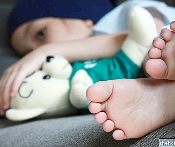
- The presence of abnormally large and deviated thumbs and big toes in such a way that they appear to move away from the other toes (medial deviation).
Pfeiffer's syndrome, therefore, is a genetic condition which, in those who carry it, mainly determines anomalies in the skull and hands.
As readers will have the opportunity to learn more in the chapter dedicated to symptoms, however, Pfeiffer's syndrome can be associated with other problems and other physical malformations.
Epidemiology: How Common is Pfeiffer's Syndrome?
According to statistics, one in every 100,000 individuals is born with Pfeiffer syndrome.
Did you know that ...
The genetic diseases that, like Pfeiffer's syndrome, cause craniosynostosis are about 150.
Among these, in addition to the Pfeiffer syndrome, the Crouzon syndrome, the Apert syndrome and the Saethre-Chotzen syndrome stand out in importance.
What causes the gene mutation associated with Pfeiffer's syndrome?
Premise: the genes present on human chromosomes are DNA sequences that have the task of producing fundamental proteins in biological processes essential to life, including cell growth and replication.
When they are free of mutations (therefore in a healthy person), the FGFR1 and FGFR2 genes produce in the right quantities, respectively, the Fibroblast Growth Factor Receptor 1 and the Fibroblast Growth Factor Receptor 2, which are two receptor proteins essential to mark the timing of cranial suture fusion and to regulate the development of the fingers and toes (in other words, they signal when it is the appropriate time for cranial suture fusion and control the formation of the fingers and feet).
On the other hand, when they undergo the mutations observed in the presence of Pfeiffer syndrome, the FGFR1 and FGFR2 genes are hyperactive and produce the aforementioned receptor proteins in such massive quantities, that the fusion times of the cranial sutures are altered (they are faster) and the process of training of the fingers and toes does not occur correctly.
Pfeiffer's syndrome is an autosomal dominant disease
To understand...
Each human gene is present in two copies, called alleles, one of maternal origin and one of paternal origin.
Pfeiffer's syndrome has all the characteristics of an autosomal dominant disease.
A genetic disease is autosomal dominant when the mutation of a single copy of the gene that causes it is sufficient to manifest itself.
Types of Pfeiffer's syndrome
In 1993, after numerous studies on Pfeiffer's syndrome, the American doctor Michael Cohen published a typological classification of the genetic disease in question, which predicted the existence of three pathological variants, identified simply with the terms "Type I", "Type II" and Type III "and all share the presence of craniosynostosis and anomalies of the thumb and big toes. The medical-scientific community immediately accepted this classification and since then the experts of Pfeiffer's syndrome have used it as a diagnostic tool and for assessing the severity of the genetic condition present; in fact, it should be noted that Dr. Cohen's classification distinguishes Pfeiffer's syndrome on the basis of the severity of the cranial and digital anomalies, and the presence of other symptoms and signs.
Going into the details of the individual pathological variants, at this point of the article it is important to underline that:
- The Type I. it is the less severe version of Pfeiffer's syndrome, as craniostenosis and thumb and big toe abnormalities have limited consequences.
Other important information: it is due to the FGFR2 mutation, sometimes combined with the FGFR1 mutation; it can be an inherited or acquired condition. - The Type II it is the most severe version of Pfeiffer's syndrome, as it is associated with severe craniosynostosis, almost incompatible with life, and with profound abnormalities in the hands and feet.
Other important information: it is exclusively due to the FGFR2 mutation; it is always an acquired condition. - The Type III it is the version of Pfeiffer's syndrome that falls, on a severity scale, just below Type II, but well above Type I, since the present craniosynostosis is almost as severe as that of the variant described in the previous point.
Other important information: it is due exclusively to the FGFR2 mutation; it is always an acquired condition.
Cranioostenosis
In carriers of Pfeiffer's syndrome, craniosynostosis can, depending on the number of cranial sutures involved in the early fusion process, have the following consequences:
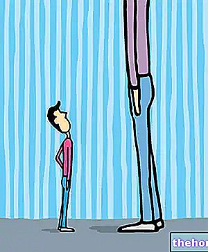
- Totally abnormal vertical development of the head, combined with a lack of lateral expansion of the skull. Thus, the patient with Pfeiffer syndrome has a long, narrow head;
- Formation of a high and prominent forehead;

- Increased intracranial pressure, on which symptoms such as persistent headache, vision problems, vomiting, irritability, hearing problems, breathing problems, mental status changes, papilledema depend;
- Intellectual deficits leading to a reduced IQ. Intellectual deficiencies are the result of the reduced space for growth enjoyed by the brain after the coronal cranial sutures have prematurely fused;
- Lack of development of the intermediate part of the face, which appears flat if not concave;
- Presence of bulging (proptosis), wide open and abnormally spaced eyes (ocular hypertelorism);
- Presence of a beaked nose;
- Failure to develop the jaw (maxillary hypoplasia), resulting in a condition of crowded teeth;
- Clover-like appearance of the head ("clover skull"). The "clover skull" causes hydrocephalus.
TYPE I
Type I Pfeiffer syndrome is associated with a mild clinical craniosynostosis, which very often is limited to giving an elongated shape to the skull and causing a visibly high forehead and a flat face.
If subjected to the right treatment, people with type I Pfeiffer syndrome usually lead a normal life and have a normal IQ.
TYPE II
Type II Pfeiffer syndrome is the only pathological variant that causes the so-called "trefoil skull", this cranial anomaly has serious repercussions on intellectual abilities and is often associated with premature death.
Those suffering from Type II Pfeiffer syndrome present the entire clinical picture described above regarding the consequences of craniosynostosis.

TYPE III
Type III Pfeiffer syndrome has the same impact on its carriers as Type II Pfeiffer syndrome, except for the "trefoil skull".
People with Type III Pfeiffer Syndrome do not enjoy a long life expectancy.
Anomalies affecting the thumbs and big toes
If particularly severe, the anomalies affecting the thumbs and big toes can profoundly compromise the functional capacity of the hands and feet, causing problems in grasping objects and / or walking.
Did you know that ...
The medial deviation affecting the thumbs and big toes of patients with Pfeiffer's syndrome is an example of varus varus. More precisely, doctors speak of thumb varus, due to the medial deviation of the thumbs, and hallux varus, due to the medial deviation of the big toes.

Brachydactyly
In Pfeiffer's syndrome, brachydactyly is a fairly common anomaly that can affect only a few fingers or the entire digital complex of the hands and / or feet.
The problem of brachydactyly can be observed in all typological variants, albeit with different frequency.
Syndactyly
In Pfeiffer's syndrome, syndactyly constitutes a fairly frequent "anomaly (less common than brachydactyly), which can have different connotations (it can be incomplete, complete, complex, etc.).
The problem of brachydactyly is observable in all typological versions of Pfeiffer's syndrome, even if with different recurrence.
Bone ankylosis
Pfeiffer's syndrome is associated, above all, with bone ankylosis of the elbow, although, in reality, it could cause the same problem to any large joint in the human body.
Bone ankylosis is a problem found only in the most severe typological versions of Pfeiffer's syndrome (especially in Type II).
Abnormalities affecting the respiratory tract
The possible anomalies in the respiratory tract induced by Pfeiffer's syndrome are such as to cause respiratory problems with serious repercussions on the general health of the patient (the brain suffers the most).
Like bone ankylosis, the above anomalies are observable only in the more severe typological variants (Type II in particular).
When is it possible to detect Pfeiffer's syndrome?
Typically, cranial and digital abnormalities due to Pfeiffer syndrome are evident at birth, so diagnosis and treatment planning are immediate.
to the head (X-rays of the head, CT scan of the head and / or MRI of the head) and hands and feet; finally, it ends with a genetic test.
Physical examination and medical history
Physical examination and anamnesis essentially consist in an accurate evaluation of the symptoms exhibited by the patient.
In the context of Pfeiffer's syndrome, it is in these phases of the diagnostic process that the doctor ascertains the craniostenosis and the anomalies affecting the thumbs and big toes, and, on the basis of the other symptoms present, hypothesizes the typological variant in progress.
Radiological examinations of the head and fingers and toes
In the context of Pfeiffer's syndrome,
- Radiological examinations of the head are used by the physician to confirm the presence of an early fusion of the cranial sutures and to estimate the severity of cranial-brain anomalies.
- Radiological examinations, on the other hand, are essential to investigate the extent of the varus and a "possible brachydactyly and / or" a possible syndactyly.
Genetic test
It is the DNA analysis aimed at detecting mutations in critical genes.
In the context of Pfeiffer's syndrome, it represents the confirmatory diagnostic test, as it allows to highlight the mutation of FGFR2 and / or FGFR1.
The genetic test is also the test that allows to establish the type of Pfeiffer syndrome present.