The main symptom of pulmonary emphysema is dyspnea, or difficulty in breathing: this, at first, appears only under exertion, then becomes manifest even at rest.

Figure: lung affected by centrilobular emphysema, characteristic of smokers. The section of the organ reveals various cavities lined with heavy deposits of black tar. From wikipedia
Diagnosis is based on imaging tests, such as chest x-rays or CT scans, and other pulmonary function tests.
Definitely recovering from pulmonary emphysema is unfortunately impossible. However, there are some treatments that can help reduce the symptoms.
Included in the list of so-called chronic obstructive bronchopneuomopathies (COPD), pulmonary emphysema represents a chronic and usually bilateral condition (ie affects both lungs).
Origin of the term emphysema. The term emphysema means "enormous dilation" or "enormous enlargement".
WHAT ARE THE ALVEOLUS?
The alveoli are small lung cavities, where gas exchanges between the blood and the atmosphere take place.


Located at the extremities of the terminal bronchioles, that is the last branches of the bronchi, the alveoli have an extended parietal surface, very elastic, which serves to increase the area for gas exchange.
Inside them, in fact, the blood is enriched with the oxygen contained in the inhaled air and it "gets rid" of the carbon dioxide produced by the tissues.
Surrounded by elastic walls, the alveoli are separated from each other by the so-called alveolar septa; these dividing structures are fundamental, because they enormously amplify the surface for gaseous exchanges, allowing better blood oxygenation.
A set of alveoli forms the so-called pulmonary acinus; the pulmonary acinus, or more simply acinus, resides at the extremity of a terminal bronchiole; the terminal bronchioles are the last branches of the lower airways, which start from the trachea and continue with the primary bronchi, the secondary bronchi, the tertiary bronchi, the bronchioles and, indeed, the terminal bronchioles.
A group of several pulmonary acini and multiple terminal bronchioles make up the smallest pulmonary structure visible to the naked eye: the lobule. In the pulmonary lobule, we can recognize more internal acini, called central, and peripheral acini, called distal.
EPIDEMIOLOGY
According to some estimates, worldwide, emphysema affects about 210 million people and causes the death of 3 million individuals every year.
At one time, it was more common among men, because the latter smoked more than women (N.B: cigarette smoking is one of the main causes of emphysema) and practiced more risky jobs.
Today, however, things have changed and, given the high number of smokers, women and men get emphysema more or less with the same frequency.
of the lungs, called Alpha 1-antitrypsin. The latter is fundamental for the good health of the alveoli, as it guarantees their elasticity and the possibility of filling up with air adequately, without damage.
But what are the alterations of the alveolar implant that give rise to emphysema?
PATHYSIOLOGY
According to the strictly medical definition, "pulmonary emphysema is:" an abnormal enlargement of the air spaces located distal to the terminal bronchiole (ie the cavities formed by the alveoli), associated with destructive lesions of the alveolar walls ".
The lesions to the alveolar walls also affect the septa that divide the various alveoli, therefore the surface for gaseous exchanges is drastically reduced. The reduction of the exchange surface is followed by less oxygenation of the blood (therefore also of the tissues) and the appearance of various respiratory problems.
Anatomically, the alveoli dilate more than normal and effectively become one.
The severity of these changes is represented by the fact that, once destroyed, the alveolar septae can no longer return to the way they were before, that is, they are irremediably damaged.
TYPES OF EMPHYSEMAS SECOND DEFINITION

Figure: Healthy alveoli and alveoli of a person with pulmonary emphysema. In the second, we can see the lack of alveolar septa and an anomalous extension of the berries. From the site: health9.org
Keeping in mind the medical definition stated above "indeed, based on the position of the affected acini," pulmonary emphysema can be divided into at least four categories:
- Centrolobular (or centroacinar) pulmonary emphysema: it presents a deterioration of the central acini of one or more lobules. It is the form of emphysema most closely related to cigarette smoking.
- Panlobular (or panacinus) pulmonary emphysema: it presents a "total alteration of one or more lobules; in other words, the terminal bronchioles, central acini and even peripheral acini are involved.
- Pulmonary paraseptal emphysema: it is due to an alteration of the peripheral pulmonary acini of one or more lobules.
- Irregular pulmonary emphysema: it presents damage to some central and some peripheral berries (which is why it is called irregular) of one or more lobules.
OTHER TYPES OF EMPHYSEMA
In fact, under the heading pulmonary emphysema it is also possible to include morbid states in which - instead of an enlargement of the alveolar spaces and a deterioration of the septa - there is an "hyperdilation or" atrophy of the lungs.
We speak of hyperdilation (or hyperdistension) in the presence of an abnormal forfeiture of air and in inadequate areas of the lungs; this condition occurs in the case of:
- Acute emphysema, typical of those suffering from asthma.
- Bullous emphysema, characterized by the formation of air bubbles.
- Interstitial emphysema, characterized by an accumulation of air around the lobules and under the pleura (lining layer of the lungs). It is usually caused by severe coughing fits.
We speak instead of atrophy of the lungs in the case of the so-called senile pulmonary emphysema. This condition is due to a shrinking of the alveoli
. Over the years, lung tissue undergoes physiological deterioration, which weakens and makes both the lungs and the alveoli more fragile.
The most characteristic clinical sign of pulmonary emphysema is dyspnea, that is, difficulty (or lack, in severe cases) of breathing.
Moments in which exertional dyspnea can appear:
- Climb the stairs
- Work that requires physical effort
- Uphill walk
- After meals
Initially, this symptom assumes the connotations of exertional dyspnea, as it arises only when the patient is engaged in physical activities that require an increase in respiratory rate.
Then, over time, the "hunger for" air becomes more severe and also appears at rest and during the most trivial tasks (dyspnea at rest).
Respiratory disorders may be associated with: cough with chronic expectoration, cyanosis (in particular in the lips and in correspondence of the nails), hyperinflation of the chest (due to an "incomplete exhalation of" inspired air), feeling of exhaustion, fever, reduced respiratory mobility (especially when the patient has to take deep breaths) and, finally, heart problems.
LUNG EMPHYSEMA: A SOMETIMES LATENT DISORDER
One of the greatest dangers of pulmonary emphysema is represented by the fact that, in some situations, the initial manifestations are almost imperceptible and remain so for several months, if not even years. This causes therapeutic treatments to start late, when the situation is already very compromised.
WHEN TO SEE THE DOCTOR?
Breathing difficulties at rest or after not particularly intense efforts should always be promptly reported to your doctor, as they could be a sign of serious respiratory and / or heart problems.
COMPLICATIONS
Pulmonary emphysema can involve the collapse of a lung due to a pneumothorax, the aggravation of heart problems and, finally, the formation of so-called "giant bubbles" in the lungs.
Going into the details:
- Pneumothorax occurs in very severe pulmonary emphysema and is due to the rupture of the acini located near the pleura, that is, the membrane that surrounds the lungs. This event, in fact, creates a passageway for the inhaled air, which, once it arrives in the lungs, exits into the adjacent pleural cavity, causing the lung to collapse.
- The aggravation of heart problems usually consists of the so-called cor pulmonale; this complication is due to the increase in pulmonary arterial pressure (ie the pressure of the blood flowing in the pulmonary artery) and is characterized by worsening of dyspnea.
- The formation of "giant bubbles", or empty spaces inside the lungs, reduces the ability of the lungs to inhale air properly. This exacerbates respiratory problems and promotes episodes of pneumothorax.
Obviously, the patient is also subjected to a physical examination, during which the doctor analyzes the extent of dyspnea and the presence of some other particular sign (cyanosis, chest inflation, etc.).
RADIOGRAPHY OF THE CHEST
Chest x-ray, or chest x-ray, is a diagnostic X-ray imaging examination, which allows the visualization of the main anatomical structures of the chest: therefore the heart, lungs, main blood vessels, most of the ribs and a portion of the spine .
The resulting images are obtained by exposing the patient to a certain dose of ionizing radiation (X-rays); in general, the information collected by chest radiography is quite clear and exhaustive, but in some particular cases of pulmonary emphysema they may present without anomalies.
CT scan
CT, or computed axial tomography, is a more sensitive imaging test than a chest X-ray, which can show the lungs from multiple angles.
Its execution, unlike the chest X-ray, allows to "find" any anomaly in the lungs and thoracic area, clarifying the exact origin of the complaints complained of by the patient.
Even the CT scan, like radiography, exposes those who undergo it to a non-negligible dose of ionizing radiation.
ARTERIAL HEMOGASANALYSIS
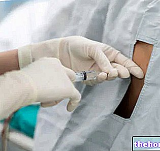
Arterial blood gas analysis is a particular diagnostic test, which takes place on a blood sample usually taken from the wrist. Through this test, the doctor measures the pressure of the gases present in the blood (therefore oxygen and carbon dioxide) and the blood pH. based on the results of the measurements, therefore, it is able to predict lung function, the efficiency of gas exchanges inside the alveoli and the oxygen levels circulating in the blood.
In the case of pulmonary emphysema, gas exchange is, as mentioned, deficient, so the blood is generally poor in oxygen.
SPIROMETRY

Figure: Spirometry. From Wikipedia
Spirometry is one of the most common and practiced diagnostic tests for estimating lung function, because it is fast, effective and painless.
During its execution, the patient must take breaths while connected via a mouthpiece to an instrument, called a spirometer; this device measures the inspiratory and expiratory capacities of the lungs and the patency (ie the opening) of the airways passing through the latter.
Spirometry, performed on a patient with pulmonary emphysema, has a characteristic outcome, which a doctor can decipher.
Pulmonary emphysema cannot be cured, as, unfortunately, the damage to the alveoli is irreparable.
However, to alleviate the symptoms and improve the quality of life, the patient can be treated with drugs, with special therapies (such as pulmonary rehabilitation and oxygen therapy) and with a specific surgery.
PHARMACOLOGICAL TREATMENTS
Based on the severity of pulmonary emphysema and associated conditions, your doctor may prescribe:
Some examples of inhaled corticosteroids:- Beclomethasone
- Flunisolide
- Budesonide
- Fluticasone
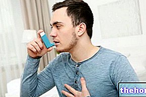
- Bronchodilators. These drugs relieve cough, wheezing and all various respiratory problems, as they improve the patency of the lower airways. Unfortunately, they are not as effective as in chronic bronchitis and asthma.
- Inhaled corticosteroids. Corticosteroids are very powerful anti-inflammatories, which are usually given when the "lighter" treatments have not worked as intended. In case of pulmonary emphysema, they are taken through aerosol sprays and are used, above all, to improve dyspnea. Their prolonged use favors osteoporosis, hypertension, the onset of diabetes and cataracts, obesity, etc. Therefore, before using them, it is advisable to consult your doctor.
- Antibiotics. The doctor may have them taken if he is concerned that the patient may get some bacterial infection, such as pneumococcal pneumonia.
OTHER THERAPIES
For the improvement of the symptoms caused by pulmonary emphysema, the following provide excellent results: respiratory rehabilitation, respiratory physiotherapy, oxygen therapy and a tailor-made diet.
Respiratory rehabilitation consists in having the patient practice a series of motor exercises (exercise bike, climbing stairs, walking, etc.), in order to improve tolerance to efforts and reduce the severity of dyspnea.
Respiratory physiotherapy aims to improve the patient's respiratory capacity, although it does not involve any strictly pulmonary benefit.
Oxygen therapy is used to increase the amount of circulating oxygen, when this, due to impaired lung function, is scarce both at the blood level and at the tissue level (ie in the body tissues).
Finally, the tailor-made diet is a nutritional measure aimed at maintaining body weight or, in the case of obesity or overweight, weight loss.
SURGICAL INTERVENTION
Surgery is used only in the case of very severe pulmonary emphysema. The operations usually provided are:
- Pulmonary reduction. It consists in the removal of the damaged parts of the lung, so that the healthy parts, left in place, can function better. It is a particularly invasive and risky procedure (post-operative mortality, after a few years, is not negligible at all. ) and a long preparation.
- Lung transplant. It is the procedure whereby the diseased lung is replaced with another healthy one, coming from a compatible donor. Given the considerable invasiveness and the fair probability of failure of the operation (rejection of the organ), lung transplantation is an operation practiced only in extreme cases and when all the other solutions mentioned above have not provided any benefit.
SOME IMPORTANT PRECAUTIONARY MEASURES
For those suffering from pulmonary emphysema, to improve the quality of their life, it is advisable:
- Stop smoking. It is also a good idea to avoid inhaling secondhand smoke, because it is just as harmful.
- Avoid places and environments in which substances irritating to the lungs circulate in the air. It is advisable to keep away from cities and polluted areas, and not to use fireplaces, stoves and wood-burning ovens in their homes.
- Practice physical activity on a regular basis. Motor exercises must, of course, be adapted to your health conditions, requiring an exaggerated effort on your lungs could be dangerous.
- Protect yourself adequately from cold air. During the winter season, it is good to repair both the mouth and the nose with a scarf, because the inhalation of cold air narrows the airways and complicates breathing.
- Preventing respiratory infections. It is of fundamental importance to resort to the anti-influenza and anti-pneumococcal (pneumonia) vaccine and to avoid any direct contact with cold and flu patients.







-nelle-carni-di-maiale.jpg)
