Generality
A meningioma is a brain tumor that originates from the meninges.
Meningiomas are often benign (non-cancerous) and rarely present with a malignant phenotype. The cause is not well understood, but some environmental and genetic factors appear to be involved in the pathogenesis. Symptoms vary depending on the site of onset, the size of the tumor mass, and whether or not other tissues or structures are involved. Treatment options include observation, surgery, and / or radiation therapy.

Cranial and spinal meninges
The meninges are protective membranes that surround and protect the brain and spinal cord. They are organized in three layers, which from the outside to the inside take the name of dura mater, arachnoid and pia mater. A meningioma arises from the arachnoid cells that form the middle layer, often appears as a slowly expanding mass, and is firmly anchored to the dura mater. Often, the tumor is localized inside the head, in particular at the base of the skull, just above the spinal cord (brain stem), around the optic nerve sheath, etc. Spinal meningiomas, which are rarer, arise instead inside the vertebral canal.
Classification and staging
Meningiomas represent a heterogeneous set of tumors.
Based on the histological characteristics, the risk of recurrence and the growth rate, the World Health Organization (WHO) has developed a classification that establishes three general degrees:
- Grade I - benign meningiomas (90%): low risk of recurrence and slow growth;
- Grade II - atypical meningiomas (7%): increased risk of recurrence and / or rapid growth;
- Grade III - anaplastic / malignant (2%): high rates of relapse and aggressive growth (invade adjacent tissues).
Causes
The causes of meningiomas are not well understood. Many cases are sporadic, that is, they arise randomly, while some respect a family transmission. Meningiomas can arise at any age, but are most commonly found between the ages of 40 and 60.
Risk factors include:
- Radiation exposure. Some patients have developed a meningioma after being exposed to radiation. Radiation therapy, particularly to the scalp, can increase the risk of developing cancer.
- Female hormones. Meningiomas occur more frequently in women, and some doctors believe that female sex hormones (estrogen and progesterone) may play a role in cancer pathogenesis. This possible link is still under investigation.
- Brain injuries: Some meningiomas have been found at the site of a previous head injury (e.g., in the vicinity of a previous fracture), but the relationship is not yet fully understood.
- Genetic predisposition. Patients with type 2 neurofibromatosis (NF-2), a rare genetic disorder that affects the nervous system, have a 50% chance of developing one or more meningiomas. Other genes could act as tumor suppressors and the lack or alteration of these would make people more susceptible to meningiomas. For example, patients with NF2 are more likely to develop meningiomas because they have inherited a gene that has the potential to induce cells. normal to neoplastic transformation. The genetic alterations most frequently found in meningiomas are inactivating mutations of the neurofibromatosis type 2 (NF2) gene on chromosome 22q. Other possible genes or loci involved include AKT1, MN1, PTEN, SMO and 1p13.
Signs and symptoms
For further information: Meningioma Symptoms
Benign meningiomas are characterized by slow progression. Small tumors (Many brain meningiomas are located just below the top of the skull or between the two hemispheres of the brain. If the tumor is in these areas, symptoms include: headache, fainting, dizziness, memory problems, and changes in the brain. of behavior.More rarely, meningioma is located near sensory areas of the brain, such as the optic nerve or near the ears; patients with these tumors can experience diplopia (double vision) and hearing loss.
The suspicion of a meningioma can also arise due to the presence of intracranial hypertension, seizures and neurological deficits.
Meningiomas that grow inward can compress, but not invade, the brain parenchyma. Outward expansion, on the other hand, can cause hyperostosis, meaning the tumor can invade and distort adjacent bone. Occasionally, a meningioma can compress blood vessels or nerve fibers. Some tumor masses contain cysts or deposits of calcified minerals, others are highly vascularized and contain hundreds of small blood vessels. Additionally, a patient may have multiple or recurrent meningiomas. The phenotype of the latter is benign, but characterized by an aggressive metabolism, similar to that observed for the atypical form.
Anaplastic / malignant meningioma is a tumor with a particularly aggressive behavior and can metastasize in the body, although, as a general rule, brain tumors do not exhibit this behavior, due to the blood-brain barrier. Anaplastic / malignant meningiomas expand within the brain cavity but tend to connect to blood vessels, so metastatic cells can enter the bloodstream. Metastases often begin in the lungs.
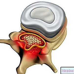
Spinal cord meningiomas are usually found in the spinal canal, between the neck and abdomen. These tumors are almost always benign and commonly manifest as pain, incontinence, episodes of partial paralysis, weakness and stiffness in the arms and legs. .
Diagnosis
The diagnostic approach involves performing some imaging tests, as is done to confirm the presence of other brain tumors. Usually, the tumor mass is found by means of magnetic resonance imaging (MRI) with contrast medium (example: gadolinium). MRI works by exposing the patient to radio waves and a magnetic field, which allow you to acquire detailed images of the brain and spine, highlighting the position and size of any tumor present.
Meningioma can also be diagnosed with computed tomography (CT). CT allows you to assess the degree of bone invasion, cerebral atrophy and hyperostosis.
Sometimes, a biopsy may be done to determine if the tumor is benign or malignant. Many people remain asymptomatic throughout their lives and are often unaware of the malignancy: in 2% of cases, meningiomas are discovered only after autopsy. With the advent of modern imaging systems, the identification of asymptomatic tumors has tripled. compared to the past.
Treatment
The best treatment for a meningioma varies from patient to patient and is established based on a number of factors, including:
- Location: If the tumor is easily accessible and causes symptoms, surgical removal is often the best treatment option.
- Size: If the tumor is less than 3 cm in diameter, stereotaxic radiosurgery may be indicated.
- Symptoms: If the tumor is small and asymptomatic, treatment can be postponed and the patient monitored with periodic neuro-imaging tests.
- General health conditions: For example, in patients with other major medical conditions, such as heart disease, there may be risks associated with general anesthesia.
- Grade: Treatment varies according to the grade of the meningioma.
- Most grade I meningiomas can be treated with surgery and continued observation.
- Surgery is the first-line treatment for grade II meningiomas. After surgery, irradiation may be required.
- Surgery is the first approach indicated for grade III meningiomas, followed by a radiotherapy regimen. If the cancer recurs, chemotherapy can be used.
Observation (growth monitoring)
Due to the slow tumor progression, medical treatment is not necessary for all patients. If a meningioma is small and is not causing symptoms, observation by magnetic resonance imaging is sufficient to periodically rule out the enlargement of the tumor mass.
Surgical excision and resection
Surgical removal is the first choice treatment for symptomatic meningiomas. If the tumor is superficial and easily accessible, the surgical approach may represent a lifelong cure. However, total resection is not always possible: some meningiomas can invade blood vessels or adjacent bone or develop near critical areas of the brain or of the spinal cord. In these cases, surgeons partially remove the tumor, removing as much of it as possible. If the meningioma cannot be completely eliminated, the remaining tissue can be treated with radiation therapy.
The necessary treatment after surgery depends on several factors:
- If no tumor remains, the patient is continuously monitored and further treatment may not be necessary;
- If not, the doctor may recommend periodic irradiation and follow-up;
- If the meningioma is atypical or malignant, a radiation therapy regimen is more likely to be indicated.
Possible risks of surgery include bleeding, infection, and damage to nearby normal brain tissue. Serious complications can include: cerebral edema (temporary accumulation of fluid in the brain), seizures and neurological deficits, such as muscle weakness, speech problems or coordination difficulties. These symptoms depend on the location of the tumor and often disappear after a few weeks. Sometimes, a second surgery is needed for patients with recurrent meningioma.
Radiotherapy
Radiation therapy can be used to combat meningiomas that cannot be removed with surgery. Irradiation is often recommended after partial resection or may be an option when the tumor is not effectively treatable with surgery.
Radiation therapy uses high-energy X-rays to reduce the tumor mass and destroy residual cancer cells. The goal of treatment is to destroy all residual cancer cells and reduce the chance of the meningioma returning. The benefits of irradiation are not immediate, but they do occur over time. Radiation therapy can stop meningioma growth and improve survival. free (i.e. prevents tumor recurrence) and global.
Radiosurgery
Stereotaxic radiosurgery is a procedure that precisely targets a single, high dose of radiation, allowing damage to surrounding healthy tissues to be minimized. Treatment has a very high probability of controlling the tumor.
Stereotaxic radiosurgery can be used for surgically inaccessible meningiomas or to treat residual cancer cells. The procedure is well tolerated and is typically associated with minimal side manifestations, such as fatigue and headache.
Conventional chemotherapy
Chemotherapy is rarely used in the management of meningioma, as surgery and radiation therapy are usually successful. However, this option is available for patients who do not respond to these treatments. The chemotherapy regimen commonly involves the use of hydroxyurea, which has been shown to slow the growth of meningioma cells.
Prognosis
The location of the tumor is the most important clinical factor in defining prognosis. The result of the surgical treatment, in fact, mainly depends on the extent of the resection / removal, which in turn is largely determined by the location of the meningioma (which can be adjacent or connected to other tissues or structures). Benign meningiomas present the higher survival rate, followed by atypical and finally malignant meningiomas. Patient age and general health conditions prior to surgery may influence outcome: younger subjects have better survival rates. In cases where the entire tumor cannot be removed, the likelihood of recurrence is greater. In the follow-up schedule, regular MRI or CT is an important part of the long-term care of any meningioma.


-nelle-carni-di-maiale.jpg)


