Generality
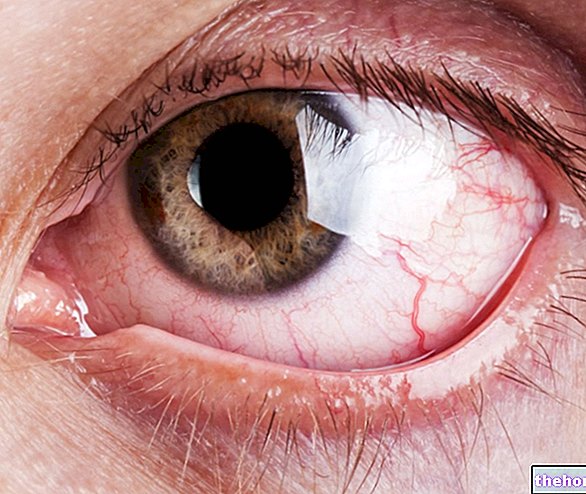
Commonly known as the "white of the eye", the sclera is the fibrous membrane that lines much of the eyeball.
Made up of dense connective tissue, this structure forms a real "shell" that stabilizes the shape of the eye, while protecting the bulbar contents.
Structure
Together with the cornea, the sclera (or sclerotic) constitutes the fibrous tunic, which is the outermost layer of the eyeball.
The sclera is mainly formed by bundles of connective tissue, containing collagen and elastic fibers, which intertwine with each other in various directions, overlapping in several layers (for comparison, the connective bundles are arranged in a similar way to the meridians and parallels of a globe). This particular "network" organization ensures mechanical resistance to the eyeball, allowing the sclerotic to perform a structural and protective function.
From a structural point of view, the sclera can be divided into 3 parts:
- episclera (very thin fibrovascular membrane, located immediately under the bulbar conjunctiva);
- sclera proper (intermediate layer consisting of consistent connective tissue);
- lamina fusca (innermost layer, leaning against the choroid).
The sclerotic has a maximum thickness of 1.5-2 mm, at the exit of the optic nerve, while it tapers in the anterior portion up to 0.3 mm.
Appearance
The sclera covers approximately the posterior 5/6 of the eyeball (in the anterior segment, the cornea occupies the remaining 1/6) and is partially visible between the eyelids.
The sclera is not a transparent anatomical structure, but is opaque and whitish. This color may decline towards blue in children (since the sclerotic membrane is thinner and the pigmentation of the underlying choroid shines through) and tends to yellowish in the elderly (mainly due to dehydration and lipid deposits).
The variation in the color of the "white part of the eye" can also depend on the presence of certain diseases. A bluish tinge due to the thinning of the sclera, for example, can occur in rheumatoid arthritis. When the eye takes on a marked color yellow, on the other hand, the cause is due to an accumulation of bile pigments (jaundice).

