What is mammography
How and why it is done
The mammography unit is used exclusively to monitor the situation of the breast and, like all machines, is subject to wear and technological developments.

Traditional mammograms use a normal X-ray film that is exposed by the passage of X-rays through the breast.
Today, on the other hand, it is possible to find mammographs that use the digital technique to obtain a more precise, safer and more reliable assessment. These devices significantly reduce the amount of radiation required, but still cost 4 to 8 times higher than traditional surveys. However, it is desirable that the spread of digital mammograms in the coming years will contribute to significantly lowering the cost of the examination.
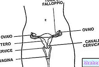
Their use is particularly useful in young women where the high density of the glandular tissue can make it difficult to read the image. In these cases, breast ultrasound examination is often recommended.
Therefore it is very important to evaluate the level of professionalism of the structure to which one is addressing. Obsolete instruments generally produce a higher radiation dose than newly designed mammograms. It is also necessary to consider the level of professionalism of the staff, since in many cases the evaluation of the images lends itself to misinterpretation.
It is important to follow your doctor's advice and to cooperate during the test. An error during the detection can in fact force a repetition of the examination by doubling, unnecessarily, the dose of radiation to which the subject is subjected.
At the end of the examination, the technician checks in advance the results which will then be subjected to the examination by the radiologist who will verify the presence of any shadows, opacities, irregular edges or anomalous masses.
Precautions
The only precaution to be implemented during preparation for the exam concerns women of childbearing age.
In these cases it is advisable to perform a mammogram in the first half of the cycle (between the end of menstruation and ovulation) since in this phase it is possible to exclude a possible pregnancy and the breasts are less tense and more compressible.
Mammography should therefore not be done during gestation, especially during the first three months as the radiation could cause serious problems to the fetus.
It is therefore advisable to avoid the examination if it is not possible to exclude a possible pregnancy with certainty.
During the postmenopausal phase, it is generally possible to carry out the investigation at any time.
Mammogram and breast size
There is no relationship between breast size and the risk of developing cancer.
Mammography is however able to identify its possible development at an early age regardless of the size of the breast since its results are influenced only by the density of the breast. The greater the presence of glandular tissue (a typical characteristic of young women), the lower is the diagnostic precision of mammography.
The size of the breast, on the other hand, influences the clinical efficacy of self-examination as it is easier to discover a lump if the breast is small.
Even a possible surgical removal operation is conditioned by the size of the breast. The choice of conservative surgery (quadrantectomy) which involves the removal of only the portion of the breast containing the neoplastic nodule, or of the total removal of the breast (mastectomy), in fact depends on both the size of the lump and the size of the breast. Obviously, with the same extension of the lump, the operation will result in greater aesthetic damage in a small breast.
Deepening: When to do a mammogram? "


.jpg)