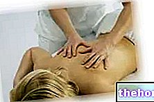
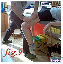
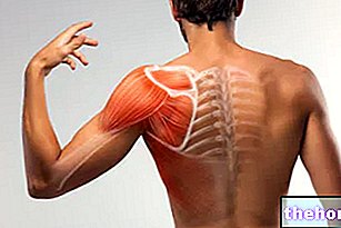
After years of working on athletes from different sports, I believe that this practice is now updated with the dynamism of the passive technique, be of great help if synergistic with the various athletic training jobs - flexibility, elasticity -for obtaining and optimizing the best Range of Motion musculo-articular. The muscles belonging to a specific myofascial group - eg those of the thigh - must be able to freely contract / decontract independently, while remaining synergistic, antagonistic or recruited during the biomechanics of an athletic gesture.

"... a well-stretched muscle, in fact, is able to perform wider movements with less stress for its joint. Studies on joint mobility confirm that performing exercises of stretching at the end of workout, accelerates recovery ... "(M. Scudero, massage therapist) in complete agreement I would like to state, as mentioned above, that to follow the word detachment when we talk about elongation / strethcing it can be decidedly appropriate and synergistic. "Furthermore, these adhesions exert both lateral traction forces, destabilizing for the associated myofascial compartment, and forces capable of establishing axial torsion on the muscles themselves" (A. Riggs). Myofascial adhesions can lead to an imbalance of the forces acting in tensegrity on a joint compartment. This imbalance generally causes postural adaptations, inflammation and pain, conditions that are detrimental to sports performance and in the most serious cases prevents the athlete from training. An example is the "gluing of the Tensor Muscle of the Fascia Lata (TFL), Ileotibial tract, with the Vasto Lateral (fig. 2).
The latter subjected to the lateral traction action exerted by the TFL, can be the cause of a knee resentment and, as some studies show, be responsible or contributing to these compartment pains as in the patellofemoral syndrome. The same TFL is capable of traction by gluing the myotendinous insertion of the biceps femoris especially during the squat phase, which can contribute to the onset of pain in the knee area due to lack of stability. In this regard it is possible to find ample documentation. in the various studies published on various Sports & Med sites, of how the detachment of the thigh and leg muscles has resolved pain and limitations for the knee joint that presented with the typical symptoms of the most common compartment syndromes such as those shown in fig. 3.

Other articles on "Passivactive technique in myofascial detachment: lower limbs - 6th part -"
- Passivactive technique in myofascial detachment: lower limbs - 5th part -
- Passivactive technique in myofascial detachment: lower limbs - 1st part -
- Passivactive technique in myofascial detachment: lower limbs - 3rd part -
- Passivactive technique in myofascial detachment: lower limbs - 2nd part -
- Passivactive technique in myofascial detachment: lower limbs - 4th part -
- Passivactive technique in myofascial detachment: lower limbs - 7th part -
- Passivactive technique in myofascial detachment: lower limbs - 8th part -
- Passivactive technique in myofascial detachment: lower limbs - 9th part -
- Passivactive technique in myofascial detachment: lower limbs - 10th part -
- Passivactive technique in myofascial detachment: lower limbs - 11th part -
- Passivactive technique in myofascial detachment: lower limbs - 12th part -
- Passivactive technique in myofascial detachment: lower limbs - 13th part -
- Passivactive technique in myofascial detachment: lower limbs - 14th part -

