Generality
Mitral insufficiency (or mitral regurgitation) is the incomplete closure of the left atrioventricular orifice, where the mitral (or mitral) valve resides; this occurs during the ventricular systole phase, that is, in the moment of contraction of the heart's ventricles; in similar conditions, finding the orifice not completely closed due to the valvular incontinence, the blood partially goes back, passing from the left ventricle to the left atrium: it is the so-called mitral regurgitation.
The causes of mitral regurgitation are numerous and such as to cause lesions in one or more components of the mitral valve. The symptoms, although less evident, are very similar to those of mitral stenosis: dyspnoea, atrial fibrillation and weakness, just to name a few.
Various instrumental methods are used to diagnose mitral insufficiency: electrocardiography, echocardiography, chest radiography and cardiac catheterization each have different advantages in assessing the extent of heart disease. Treatment depends on the severity of the mitral regurgitation: if the situation is critical, surgery is required.
What is mitral insufficiency
Pathological anatomy and pathophysiology
Mitral regurgitation, also called mitral regurgitation, consists in the incomplete closure of the left atrioventricular orifice, presided over by the mitral (or mitral) valve.
Under normal conditions, during ventricular systole (when the ventricle contracts), the mitral valve hermetically closes the passage between the atrium and the ventricle; consequently the blood flow takes only one direction, towards the aorta.
In the presence of mitral insufficiency, the pathological event occurs precisely during the ventricular systole phase: when the ventricle contracts, a portion of blood, instead of entering the aorta, goes back and goes up to the left atrium above. For this reason , mitral regurgitation is also called mitral regurgitation.
Before examining what a mitral valve looks like and how it works in cases of mitral insufficiency (analyzing its pathological anatomy and pathophysiology, respectively), it is useful to mention some fundamental characteristics of the valve:
- The valve ring. Circumferential structure of connective tissue that delimits the valve orifice.
- The valve orifice measures 30 mm in diameter and has an area of 4 cm2.
- Two flaps, front and back. For this reason, the mitral valve is said to be bicuspid. Both flaps enter the valve ring and face the ventricular cavity. The anterior flap faces the aortic orifice; the posterior flap, on the other hand, faces the wall of the left ventricle. The flaps are composed of connective tissue, rich in elastic fibers and collagen. To facilitate the closure of the orifice, the edges of the flaps possess particular anatomical structures, called commissures. There are no direct controls, of a nervous or muscular type, on the flaps. Likewise, there is no vascularization.
- The papillary muscles. There are two of them and they are extensions of the ventricular muscles. They are supplied by the coronary arteries and give stability to the tendon cords.
-
The tendon cords. They serve to join the valve flaps with the papillary muscles. As the rods of an umbrella prevent it from turning outward in strong winds, the tendon cords prevent the valve from being pushed into the atrium during ventricular systole.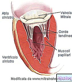
In the presence of "mitral insufficiency, on the basis of the triggering cause, lesions are created to one or more of these components of the valve. On the basis of the effects induced by each cause, two types of mitral insufficiency are distinguished, each of which groups different physiopathological behaviors. We therefore have:
- Acute mitral insufficiency.
- Chronic mitral insufficiency.
The difference between the acute and the chronic form depends, first of all, on the rapidity with which the heart disease itself is established. Before deepening this point, however, it is necessary to clarify some pathophysiological aspects common to both forms.
In the case of mitral insufficiency, both the left atrium and the left ventricle affect the pathological adaptation of the blood flow. Under normal conditions, during ventricular systole, the hermetic closure of the mitral ensures unidirectional blood flow towards the aorta. In the presence of "mitral insufficiency, however, the left ventricle pumps blood in two directions: aorta (correct direction) and left atrium (wrong direction due to" valvular incontinence). Therefore, the amount of blood that reaches the tissues is reduced and its flow varies according to the size of the orifice: the less efficient the mitral closure, the greater the amount of blood that returns to the atrium (regurgitated fraction) and the lower the cardiac output. The left atrium also dilates accordingly to accommodate the greater amounts of blood.
During diastole, ie in the relaxation phase of the ventricles and atria, the regurgitated blood (in the atrium) returns to the ventricle, as the mitral valve opens in this phase.
This last abnormal movement of blood and the previous regurgitation have effects on the atrioventricular pressure gradient. By gradient we mean a variation, in this case of pressure. In fact, in the presence of a mitral stenosis the pressure relationship, existing between the two compartments, changes from normal The pressure changes are due to the amount of blood regurgitated, which, stopping first in the atrium and then in the ventricle, is added to that coming from normal circulation. This happens at the wrong times and it all results in an increase in ventricular pressure. In this case, we speak of left ventricular decompensation.
If the cause of mitral insufficiency slowly determines this scenario just described, the left ventricle manages to adapt to the changes (chronic form): it becomes hypertrophic, in such a way as to keep the increase in pressure inside it under control. the hypertrophic ventricular walls, at the moment of contraction, counterbalance the considerable tension caused by the high pressure and the regurgitated portion remains stable. This situation, however, creates a slow deterioration of the ventricular walls, destined to result in a decrease in cardiac output.
If the cause of mitral insufficiency, on the other hand, rapidly develops the pathophysiological mechanisms described above, the left ventricle does not have sufficient time to adapt to the change and does not become hypertrophic (acute form). The walls of the ventricle are therefore unable to withstand the tension caused by the high pressure and the extent of the regurgitation of blood progressively increases. This causes a continuous increase in pressure inside the left atrium, such as to affect the vessels and districts located upstream, the pulmonary veins and the lungs, with the possible development of edema.
Causes
The causes of mitral regurgitation are numerous. Each of them causes lesions of one or more structural elements that make up the mitral valve; sometimes, it can happen that two different causes, when added together, result in a lesion of a single valve component.
In the case of acute mitral regurgitation:
Alterations of the mitral ring
Alterations of the valve leaflets
Rupture of the tendon cords
Alterations of the papillary muscles
Infective endocarditis; trauma; acute rheumatic disease; idiopathic; myxomatosis degeneration (collagenopathy); coronary heart disease; malfunction of the valve prosthesis.
In the case of chronic mitral insufficiency:
Alterations of the mitral ring
Alterations of the valve leaflets
Rupture of the tendon cords
Alterations of the papillary muscles
Inflammatory; rheumatic heart disease; calcification; myxomatosis degeneration (collagenopathy); infectious endocarditis; cardiac ischemia; Marfan syndrome (congenital); valve fissure (congenital); mitral valve prolapse (congenital); connect.
The two forms of mitral regurgitation therefore share only a few causes.
Symptoms and signs
The main symptomatology of mitral insufficiency, although less obvious, has numerous similarities with that which characterizes mitral stenosis.
- Dyspnea from exertion.
- Heartbeat (palpitation).
- Respiratory infections.
- Asthenia.
- Chest pain, due to angina pectoris.
- Pulmonary edema.
Exercise dyspnea is difficulty breathing. In the specific case, it arises from the decreased cardiac output of the left ventricle, due to the amount of blood regurgitated towards the atrium. Therefore, the body's response consists in increasing the number of respiratory acts, in order to counterbalance the reduced supply of oxygen due to the insufficient volume of the throw.
Pulmonary edema is a typical symptom of acute mitral insufficiency. The rapid onset of heart disease does not allow the ventricle to limit the effects induced by the increase in ventricular pressure. Unlike what happens in forms of chronic insufficiency, the left ventricle, in fact, does not have time to become hypertrophic. Consequently the altitude of regurgitated blood progressively increases, which results in an increase in pressure, not only in the left atrium, but also in the vessels and districts located upstream, ie pulmonary veins and lungs. The increased pulmonary pressure (pulmonary hypertension) causes compression of the respiratory tract and, in the most serious cases, the leakage of fluids from the vessels to the alveoli. This last condition is the prelude to pulmonary edema: in these conditions, oxygen exchange - carbon dioxide between alveoli and blood is compromised.
Heartbeat, also known as palpitation, is the most common symptom of mitral regurgitation. It consists of an increase in the intensity and frequency of the heartbeat. In this specific case, heartbeat can result from atrial fibrillation
Atrial fibrillation is a "cardiac arrhythmia, that is, an alteration of the normal heartbeat rhythm." It is due to a disorder of the nerve impulse coming from the sinoatrial node. It results in fragmentary and hemodynamically ineffective atrial contractions (that is, what concerns the blood flow).
In the case of mitral regurgitation, the regurgitation of blood in the atrium alters the volume of blood pushed into the aorta by ventricular contraction. In light of this, the body's oxygen demands are no longer met. Faced with this situation, the individual, affected by atrial fibrillation, increases breathing, palpitations, pulse irregularities and, in some cases, fainting due to lack of air. The picture can degenerate further: a continuously increasing regurgitation and the accumulation of blood in the vascular systems upstream of the left atrium, if associated with an impaired coagulation, give rise to the formation of thrombus (solid, non-mobile masses, composed of platelets ) inside the vessels. The blood clots can break down and release particles, called emboli, which, traveling through the vascular system, can reach the brain or heart. In these locations, they become an obstacle to the normal circulation and oxygenation of brain or cardiac tissues, causing the so-called ischemic stroke (cerebral or cardiac). In the case of the heart, it is also referred to as a heart attack.
Unlike what happens for mitral stenosis, embolisms due to mitral insufficiency are rarer.
Respiratory or thoracic infections are due to pulmonary edema.
Chest pain due to angina pectoris is a rare event. Angina pectoris is due to left ventricular hypertrophy, ie of the left ventricle. In fact, the hypertrophic myocardium needs more oxygen, but this request is not adequately supported by the coronary implant. It is therefore not the consequence of an obstruction of the coronary vessels, but of an imbalance between the consumption and the supply of oxygen to the tissues. .
The characteristic clinical sign of a "mitral insufficiency is the systolic murmur. It originates from the regurgitation of blood, through the half-open valve, during the ventricular systolic contraction.
Diagnosis
Mitral regurgitation can be detected by the following diagnostic tests:
- Stethoscopy.
- Electrocardiogram (ECG).
- Echocardiography.
- Chest x-ray.
- Cardiac catheterization.
Stethoscopy. Detection of a systolic murmur is the most useful clue to diagnosing mitral valve insufficiency. The murmur is produced when the regurgitation of blood passes from the left ventricle to the left atrium. It is perceived in the systolic phase, since it is at this moment that the mitral valve is not closed as it should. A strong murmur is indicative of "moderate insufficiency, but not necessarily a strong one. In fact, a weak murmur is perceived both in individuals with mild mitral insufficiency and in subjects with severe (ie severe) insufficiency. The latter" situation is the consequence of a progressive degeneration of the left ventricle. The detection zone is in the 5th intercostal space, that is, the one coinciding with the position of the mitral valve.
ECG. By measuring the electrical activity of a heart with mitral insufficiency, the ECG shows:
- Hypertrophy of the left ventricle.
- Left atrium overload.
- Atrial fibrillation.
- Cardiac ischemia.
Diagnosis by ECG gives an idea of the degree of severity of mitral insufficiency: if the outcome is comparable to that of a healthy individual, it means that it is not a severe form; vice versa, the examination shows the aforementioned irregularities.
Echocardiography. Using the ultrasound emission, this diagnostic tool shows, in a non-invasive way, the fundamental elements of the heart: atria, ventricles, valves and surrounding structures. From echocardiography, the doctor can detect:
- Abnormal behavior of the flaps, due to injury to the valve tendon cords.
- Anomalies of the left ventricle, during the phases of systole and diastole.
- Increased size of the left atrium (dilated atrium).
- The maximum flow rate and turbulent systolic flow of regurgitation, using continuous and pulsed Doppler techniques, respectively. From the first measurement, the pressure gradient between the left atrium and the left ventricle can be obtained; from the second, the extent of the regurgitation.
Chest X-ray. It is useful for observing the situation in the lungs, verifying whether or not edema is present. In addition, it allows you to see the typical pathological changes:
- Left atrium dilated by the regurgitation of blood.
- Hypertrophic left ventricle.
- Calcification, determined by particular causes, of the valve or of the ring.
Cardiac catheterization. It is an invasive hemodynamic technique. A catheter is introduced into the vascular system and brought to the heart. Inside the vascular and cardiac cavities, it acts as an investigating probe. The purposes of this examination are as follows:
- Confirm the clinical diagnosis.
- To evaluate in quantitative terms the haemodynamic alterations, that is of the blood flow in the heart vessels and cavities.In particular, the lung condition is explored.
- Confidently define whether surgery can be performed.
- Evaluate the possible presence of other valve dysfunctions.
Therapy
The therapeutic approach varies according to the severity of the mitral regurgitation. The mild, asymptomatic forms require preventive measures to avoid bacterial infections, such as endocarditis, which affect the heart cavities.
The first appearance of symptoms and moderate / severe forms require more attention, through drug therapy and, possibly, surgery.
The most used drugs, in symptomatic cases of mitral insufficiency, are:
- ACE inhibitors. They are inhibitors of the enzymatic system that converts angiotensin. They are hypotensive drugs, which reduce the increased pressure inside the left atrioventricular cavities and the vascular systems that reside upstream.
- Diuretics. They are also hypotensive.
- Vasodilators. Example: nitroprusside.
- Digital. It is used for atrial fibrillation.
Surgery becomes essential in some critical situations: when the patient has a severe form of chronic mitral insufficiency, or when he is afflicted by an acute form.
There are two possible surgical operations:
- Replacing the valve with a prosthesis. It is the most used intervention for the valves of those individuals, not young, with serious anatomical anomalies. A thoracotomy is performed and the patient is placed in extracorporeal circulation (CEC). The extracorporeal circulation is implemented through a biomedical device which consists in creating a cardio-pulmonary pathway replacing the natural one. In this way, the patient is guaranteed an artificial and temporary blood circulation that allows surgeons to interrupt the flow of blood in the heart, diverting it to another equally effective path; at the same time, it allows to operate freely on the valve apparatus. Prostheses can be mechanical or biological. Mechanical prostheses require, in parallel, an anticoagulant drug therapy. Biological implants last 10-15 years.
- Mitral valve repair. It is an approach indicated for mitral insufficiencies due to modifications of the valve structures: ring, cusps, tendon cords and papillary muscles. The surgeon acts differently, based on where the valve lesion resides. Again, patients are placed in extracorporeal circulation. It is an advantageous technique, as prostheses have drawbacks: as we have seen, the biological ones must be replaced after about 10-15 years, while the mechanical ones require the continuous administration, in parallel, of anticoagulants. It is a method that is not suitable for rheumatic forms of mitral insufficiency: these, however, are rare.


