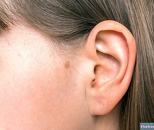
Covered with skin and consisting mainly of elastic cartilage, the auricle is a structure with a particular shape, on which anatomists recognize various characteristic areas that they call: helix, antelice, tragus, antitragus, scaphoid fossa, basin, intertragic notch and ear lobe .
In addition to the cartilage component, the auricle also includes muscles, ligaments and a small portion of adipose tissue.
The pinna can suffer from a condition known as auricular perichondritis and can be the subject of congenital or acquired malformations.
The task of the external ear is to locate, receive and amplify the acoustic signal and, subsequently, lead it to the eardrum, the first of the various structures making up the middle ear.
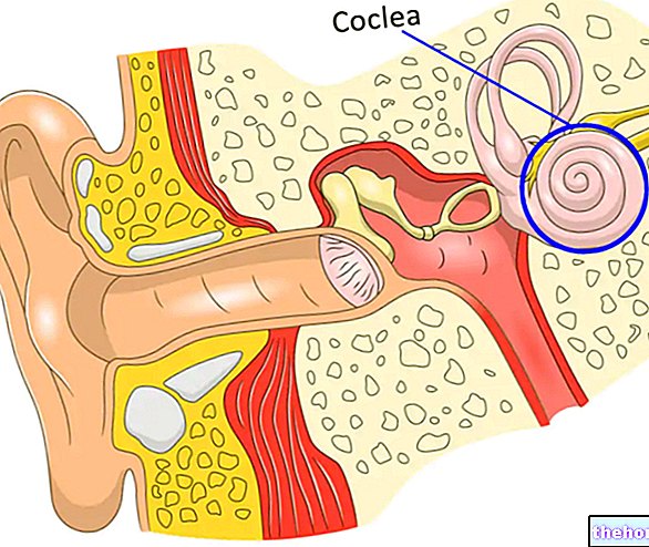
Partly visible to the naked eye, the outer ear is made up of the pinna and external auditory canal (or external acoustic meatus), and includes a number of muscles, ligaments, nerves and blood vessels.
For further information: External Ear: How it is Done and How It Works ; the temporal bone is the even and symmetrical bone of the latero-inferior region of the cranial vault, placed to protect the temporal lobe of the brain and the middle and inner ear.



.jpg)


