Symptoms, signs and complications
For further information: Hydronephrosis Symptoms
The symptoms of a hydronephrosis depend on several characteristics and, precisely, vary according to three parameters:
- Speed at which the urinary tract is blocked
- Degree of closure, partial or total
- Unilateral or bilateral obstruction
It is quite understandable how the second and third parameters affect the symptoms. A total closure of the urinary tract, in fact, is much more serious than a partial closure; the same goes for bilateral hydronephrosis: in fact, the involvement of both kidneys implies a superior renal malfunction, compared to when hydronephrosis is unilateral.
The first parameter, however, deserves a separate discussion. Depending on how quickly the passage to the urine stream closes, there can be two different forms of hydronephrosis, in some respects similar and in others different.
When the occlusion forms rapidly, we speak of acute hydronephrosis; when, on the contrary, the obstruction is created slowly, we speak of chronic hydronephrosis.
ACUTE HYDRONEPHROSIS
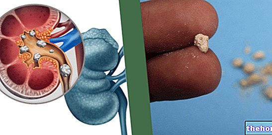
Acute hydronephrosis is usually due to kidney stones and develops in a few hours (which is why it is called acute).
The main symptom of acute hydronephrosis is severe pain, occurring in the lower back and one or both hips (between the ribs and hip). Sometimes, the painful sensation can also be felt in the testicles (in men) or in the vagina (in women). It also tends to come and go and get worse when the patient drinks something.
The following symptoms and signs are added to the pain:
- Nausea and vomit
- Difficulty urinating
- Bacterial kidney infections, due to urine stagnation
- High fever, above 38 ° C
- Uncontrollable chills
- Blood in the urine (irrefutable evidence of kidney stones)
- Swelling of the kidneys when hydronephrosis is severe
CHRONIC HYDRONEPHROSIS
The condition of chronic hydronephrosis, on the other hand, sets in very slowly: in fact, weeks, if not months, are required. The explanation of this graduality is almost certainly to be linked to the causes: for example, a tumor in the reproductive organs, pregnancy etc. are slow processes which take time to cause hydronephrosis.
Symptoms and signs do not differ much from those described for acute hydronephrosis. Pain is always the main manifestation of the disorder, with the only difference that, in some cases, it is lighter.
WHEN TO SEE THE DOCTOR?
It is advisable to contact your doctor promptly when you experience severe pain in your side (or back) and, at the same time, difficulty urinating and high fever. The latter is very often synonymous with a bacterial kidney infection, which, if not treated, can cause complications.
COMPLICATIONS
Complications usually arise in the presence of severe "hydronephrosis that is not treated promptly and appropriately.
- Tiredness
- Swelling in the ankles and hands, due to water retention
- Shortness of breath
- Malaise
- Blood in the urine
The greatest dangers faced by patients with hydronephrosis are renal atrophy and renal failure. These two pathological conditions are more likely to occur when hydronephrosis is bilateral, because in the unilateral forms the healthy kidney is able to compensate for the diseased one.
Another danger to avoid, which can arise as a result of a poorly treated hydronephrosis, is sepsis. Sepsis occurs when bacterial kidney infection, resulting from the stagnation of urine, also affects the blood.
Diagnosis
As with many diseases, a pre-diagnosis of hydronephrosis is established by the doctor thanks to a careful physical examination. Subsequently, for a certain diagnosis, an ultrasound and further examinations, both clinical and instrumental, are required to identify the causes and any associated pathologies and complications.
OBJECTIVE EXAMINATION
The doctor asks the patient about the symptoms felt and tries to identify some specific signs of hydronephrosis. Based on the description, it is also possible to get an idea of the type of hydronephrosis and its causes; for example, if the onset of symptoms was sudden (acute form), the patient probably also suffers from kidney stones; vice versa, if the symptoms started gradually, at the origin there may be a tumor or a benign enlargement of the prostate.
ULTRASOUND
Ultrasound is the instrumental examination that allows us to establish whether the patient is suffering from hydronephrosis or not. Through the ultrasound images, in fact, the specialist doctor is able to see the kidney swelling, due to the accumulation of urine.
CLINICAL TESTS
By performing some clinical tests, it is possible to clarify part of the causes and find out if the hydronephrosis has caused complications.
- Blood tests. They are used to assess whether there is "a" bacterial infection in progress.
Furthermore, they provide a quantitative datum of the creatinine values present in the bloodstream. It should be noted that this last element is not always reliable, since, especially in unilateral hydronephrosis, it could appear normal. The reason is linked to the fact that the healthy kidney compensates for the deficiencies of the diseased one, thus leaving the creatinine levels in the blood unaltered. . - Urine tests. They are used to assess if there are traces of blood. In fact, their presence is almost always synonymous with kidney stones.
OTHER INSTRUMENTAL EXAMINATIONS

Figure: Contrast urography of a patient with hydronephrosis. The left kidney (with respect to the reader) is the one affected by the disease. The contrast medium shows dilation of the renal pelvis (or pelvis) and ureter. From the website: doctorshangout.com
Once the diagnosis of hydronephrosis has been established, in order to set up a correct therapy it is very important to shed light on some details of the disorder, such as where the obstruction is or whether there is a tumor at its origin. Excellent information is obtained from:
- Intravenous urography. It is a radiological examination, which allows to evaluate the urinary tract from a morphological and functional point of view. It involves the use of X-rays and a contrast medium (iodized solution), injected into the bloodstream. The contrast medium, highlighting the urinary flow through the kidneys, ureters, bladder and urethra, allows to identify where the obstruction resides.
- CT (computed tomography). It provides three-dimensional images, relating to the internal organs of the human body. If there are tumors at the origin of the hydronephrosis, they are traced. CT scan uses ionizing radiation and is therefore considered a moderately invasive test.
DIAGNOSIS OF FETAL HYDRONEPHROSIS
Fetal hydronephrosis is recognized through routine ultrasound scans the mother undergoes during pregnancy.
Treatment
The choice of the most appropriate treatment depends on the causes and the severity of the hydronephrosis itself.
In most cases, surgery is needed. The first step is to remove the accumulation of urine in the kidney, to avoid the onset of an infection, the second step is to remove the obstruction of the urinary tract; the third definitive step is dedicated to treating the causes, such as kidney stones or a possible tumor of the reproductive organs.
The sooner you intervene, the better the patient will benefit from it. This indication is valid for both unilateral and bilateral hydronephrosis.
Untreated hydronephrosis, as we have seen, can cause sepsis and severe kidney damage (atrophy and kidney failure).
In summary, the objectives of hydronephrosis therapy are:
- Remove the stagnation of urine and the enormous pressure it causes inside the kidney involved (the swelling)
- Prevent renal atrophy and the spread of bacterial infections
- Remove the occlusion in the urinary tract
- Treat the causes of hydronephrosis with the most appropriate treatments
THE DRAINAGE OF URINE
The drainage of the urine, accumulated in the renal pelvis, is essential to reduce the pressure inside the kidney and to relieve the painful sensation. It also prevents the spread of bacterial infections, first in the renal organ, and then in the blood (sepsis).
There are at least two urinary drainage techniques and their application depends on where the obstruction is.
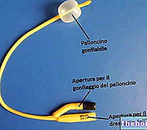
- Bladder urinary catheter. This practice involves the insertion, through the urethra, of a catheter inside the bladder. Through the catheter, the urine is drained, especially when the occlusion is in the final tract of the urinary tract.
- Nephrostomy. It consists of inserting a small tube into the kidney, specifically in the renal pelvis. The tube is inserted through a small skin incision and allows the drainage of stagnated urine.
THE SURGICAL TREATMENT: THE URETERAL STENT
Once the drainage has been carried out, it is necessary to allow the urine to flow normally again within the urinary tract.
To do this, the surgical technique used is called a ureteral stent. The ureteral stent consists of inserting a small tube inside the occluded ureter.
It serves to keep the canal open, thus allowing urine to pass through the involved urinary duct again.
Main side effects of ureteral stent:
- Dislocation of the tube
- Urinary tract infections
- Injury of the ureter and consequent scarring
- Incontinence
- Kidney pain
Usually, the ureteral stent is a temporary measure, waiting for the causes of hydronephrosis to resolve (kidney stones, tumors, etc.).
This is an effective procedure, but it can cause complications for some.
TREATMENT OF THE CAUSE
After drainage and removal of the obstruction, the third fundamental step is to treat the causes. Therefore, at this juncture, the therapeutic choice varies from patient to patient.
In presence of:
- Kidney stones: the so-called bombardment or lithotripsy is used.
- Prostatic hyperplasia: drugs are used (especially if benign) and, in more serious situations (malignant tumor), surgical removal of the prostate is used.
- Tumors of the reproductive organs: chemotherapy, radiotherapy and surgical removal of tumor tissues are needed.
HYDRONEPHROSIS IN PREGNANCY
At the end of pregnancy, hydronephrosis resolves spontaneously, therefore it is not necessary to intervene in an invasive way, if not periodically practicing the drainage of urine.
FETAL HYDRONEPHROSIS
Most cases of fetal hydronephrosis do not require specific treatments. In fact, kidney disease almost always resolves at birth or within a short time.
If the origin of "hydronephrosis c" is the so-called primary vesicoureteral reflux, two countermeasures may be required: antibiotics, against possible kidney infections, and the injection of a particular liquid substance, to prevent reflux.
Fetal hydronephrosis rarely requires surgical intervention for its resolution; surgery which, when necessary, is very similar to that of adults.
Prognosis and prevention
The prognosis is variable, as it depends on numerous factors, such as:
- Causes of Hydronephrosis
- Involvement of one or both kidneys
- Severity of the obstruction blocking the urinary tract
- Form of hydronephrosis, acute or chronic
- When you have intervened with the appropriate care
To clarify what has just been said, it is useful to give some examples.
For a patient with kidney stone hydronephrosis, the prognosis is positive, as kidney stones are effectively treated nowadays. Conversely, for an individual with hydronephrosis from malignant prostate cancer, the prognosis will be much worse. , although there are also appropriate treatments.
Timeliness and rapidity of action are fundamental: intervening at the beginning of the disorder means anticipating and preventing complications (kidney infections, sepsis, renal atrophy, renal failure, etc.); on the contrary, delaying the diagnosis and "therapeutic intervention means reducing the" efficiency of treatment and consequently worsen the prognosis.
PREVENTION
A balanced diet prevents kidney stones, and consequently kidney ailments as well. The same goes for tumors: a healthy lifestyle, in general, removes the risk of neoplasms and their possible complications. All this can serve to prevent hydronephrosis, with excellent results.

