Generality
Urinary incontinence is involuntary loss of urine. The disorder can result from a variety of conditions, including physical damage, aging, cancer, urinary tract infections and neurological disorders. Some of these causes involve only temporary and easily treatable discomfort. while other problems are more serious and persistent.
Urinary incontinence can have a profound impact on the patient's emotional, psychological, and social well-being. However, it almost always results from an underlying medical condition that can be successfully managed or treated.

The clinical picture that characterizes the inability to control the emptying of the bladder is called enuresis.
Often, the term enuresis is used in reference to urinary incontinence in children, due to a delay in the acquisition of full ability to control urination; for example, nocturnal enuresis (bedwetting) is typical. On the other hand, we tend to speak of urinary incontinence in reference to adults who, for one reason or another, lose this ability to control after having normally acquired it as children.
Note. Urinary incontinence is a common symptom of many health problems.
what happens under normal conditions?
Urinary function is controlled by a "synergistic activity between the urinary tract and the brain. In particular, continence and urination imply a balance between voluntary muscular actions (somatic nervous system) and involuntary ones (regulated by the autonomic nervous system and coordinated by a reflex mechanism).
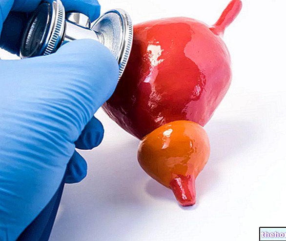
When the urination is completed, the filling phase begins: the urine is collected in the bladder, where it accumulates until the moment of its elimination, which occurs through the urethra. The bladder performs a function both as a reservoir (accumulation of urine) and as a pump (expulsion of urine).

As soon as social conditions permit - with the bladder neck open and the detrusor muscle compressing the bladder - urine flows into the urethra and the person consciously relaxes the external urethral sphincter muscles to urinate. This decision is voluntary, so during urination the urinary flow can be voluntarily interrupted with the contraction of the external sphincter. The will to retain urine has a limit, however, and if the urination reflex is sufficiently intense (due to an abnormal stretching of the bladder walls) the reflex inhibition of the external sphincter prevails over the voluntary commands that oppose urination.
Continence, both in men and women, is therefore entrusted to the presence of two main sphincters, one proximal (at the level of the bladder neck, not controlled by the will), and one distal located at the level of the urethra (under the control of the voluntary nervous system). The pelvic muscles and the ligaments that support the bladder neck and urethra, as well as all the nervous structures involved, also participate in continence.
Incontinence occurs if bladder neck closure is insufficient (stress incontinence) or if the muscles surrounding the bladder are overactive and contract involuntarily and suddenly (urge incontinence).
Causes
The disorder is more common in the female population, both for the anatomy of the urinary tract and for hormonal implications.
Several scientific studies have found that pregnancy and childbirth (via caesarean section or vaginal delivery) can increase the risk of urinary incontinence. In such cases, a weakening of the muscles and ligaments of the pelvic floor occurs, which causes a condition called urethral hypermobility (the urethra does not close properly). Urinary incontinence affects about 20-40% of women after childbirth; most of the time it is transient (it disappears spontaneously within a month or so) and, as we will see later, it is mostly "due to exertion". Prolapse too. of the uterus can cause incontinence. This condition occurs in about half of all women who have given birth. During menopause, female subjects may experience urine leakage due to decreased estrogen levels and it is interesting to note that estrogen replacement therapy has not been shown to be helpful in symptom management.
Men tend to experience urinary incontinence less often than women. Benign prostatic hyperplasia (enlarged prostate gland) is the most common cause of urinary incontinence in men over the age of 40. Prostate cancer and certain medical treatments for its management are sometimes associated with the disorder. The outcome of surgery or radiation therapy, for example, can damage or weaken the muscles that control urination.
In men and women, the aging process causes a general weakening of the urethral sphincter muscles and a decrease in bladder capacity.
Some cases of urinary incontinence are temporary and are often caused by lifestyle. Drinking alcohol, caffeinated beverages, or any liquid in excessive amounts can cause a loss of bladder control. Certain drugs can also induce a short period of incontinence: diuretics, estrogens, benzodiazepines, antidepressants and laxatives. In addition, some health conditions are associated with the disorder: diabetes, hypertension, back problems, obesity and Alzheimer's disease. Constipation and urinary tract infections can increase the need to urinate. Disorders such as multiple sclerosis, spina bifida, Parkinson's disease, stroke, and spinal cord injuries can also interfere with nerve function in the bladder.
Possible conditions that contribute to and / or cause urinary incontinence
- Vaginal or urinary tract infections
- Kidney disease;
- Pregnancy and childbirth;
- Constipation;
- Medicines;
- Diabetes;
- Enlarged prostate (benign hyperplasia) and prostatitis (inflammation of the prostate gland)
- Diseases of the nervous system and neurological disorders (for example: multiple sclerosis, Parkinson's disease, spinal cord injury and stroke);
- Congenital defects (present at birth);
- Certain surgical procedures (nerve or muscle damage)
- Weakness of the muscles that hold the bladder and urethral sphincter in place.
Types of urinary incontinence
Stress urinary incontinence
Also known as stress urinary incontinence, it is essentially caused by the loss of support of the urethra which is usually the result of damage to the pelvic floor muscles from childbirth or other causes.

Urge urinary incontinence
This type of incontinence is accompanied by a sudden and strong urge to urinate, which does not leave enough time to reach the bathroom (inability to inhibit, block or delay the urge to urinate). Urge incontinence is caused by improper (uninhibited) contractions of the detrusor muscle during the filling phase and is characterized by the leakage of large amounts of urine. When this occurs, the urge to urinate cannot be voluntarily suppressed. Risk factors for urge incontinence they include aging, obstructed urine flow, inconsistent emptying of the bladder and a diet rich in irritants (such as coffee, tea, cola, chocolate, and acidic fruit juices).
Mixed urinary incontinence
It is a combination of urge and stress incontinence.
Regurgitation urinary incontinence
It occurs when the bladder does not empty completely, if there is an obstacle to normal urine flow, or if the destrusor muscle cannot contract effectively. It is characterized by post-voiding drip (a phenomenon in which the bladder slowly leaks residual urine into the urethra after emptying). Causes of regurgitation urinary incontinence include: tumors, constipation, benign prostatic hyperplasia, and nerve damage. Diabetes, multiple sclerosis and shingles can also cause this problem.
Structural incontinence
Rarely, congenital structural problems can cause incontinence, usually diagnosed in childhood (example: ectopic ureter, posterior urethral valves, exstrophy-epispadias complex). Vesico-vaginal and uretero-vaginal fistulas, caused by trauma or gynecological injury, can lead to urinary incontinence.
Functional incontinence
It can also occur in the absence of a biological or medical problem. Patients with functional incontinence have mental or physical disabilities, which prevent them from urinating normally, even if the urinary system itself is structurally intact. The person recognizes the need to urinate, but cannot or does not want to go to a toilet. As we have seen, beyond a certain threshold of bladder filling, the involuntary reflex of urination overcomes the voluntary control of the same → the loss of urine can therefore be high. Conditions that can lead to functional incontinence include: Parkinson's disease, Alzheimer's, mobility disorders, alcohol abuse, reluctance to use toilets due to severe depression or anxiety, mental confusion and dementia.
Transient incontinence
It occurs temporarily and can be triggered by drugs, adrenal insufficiency, mental retardation, reduced mobility and severe constipation.
Diagnosis
As with any health problem, a "careful medical history and a thorough physical examination are essential. A urologist, first, can ask the patient questions about individual habits and can gather information related to personal and family medical history. loss of voiding control suggests the type of incontinence being faced.
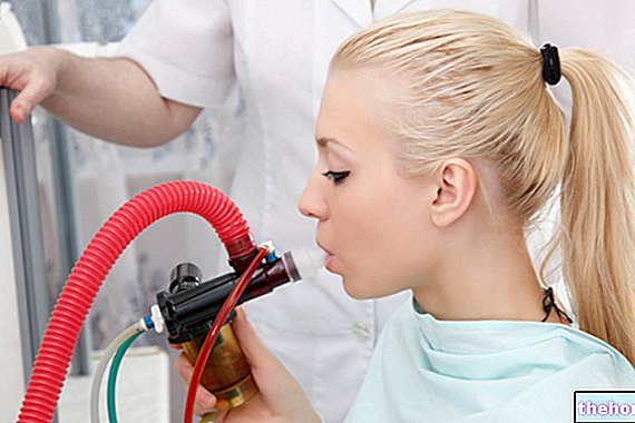
The physical exam focuses on looking for signs of particular medical conditions that cause incontinence, including constipation, prolapse, hernias, urinary tract obstruction, and neurological disorders. Usually, blood and urine tests are performed at the first evaluation to find evidence of infection, urinary stones or other causes contributing to urinary incontinence. If results suggest that further evaluation is needed, investigations such as cystoscopy or urodynamics, performed to measure bladder capacity, urine flow, and post void residue, may be recommended, as well as establishing pelvic muscle malfunction. .
Treatment
Treatment for urinary incontinence depends on the type of incontinence, the severity of the problem, the underlying cause, and what measures are best suited to the patient's lifestyle. Additionally, some treatment approaches are optimal for men, while others are more suitable for women. The goal of any urinary incontinence treatment is to improve the patient's quality of life. In most cases, the first line of treatment is conservative or minimally invasive. Medication may be needed depending on the cause of the incontinence. If symptoms are more severe and all other treatments are ineffective, a surgical approach may be recommended. Therapeutic success depends primarily on correct diagnosis. part of the cases, it is possible to achieve great improvements and resolution of symptoms.
Conservative treatments
- Lifestyle changes: Significant weight gain can weaken pelvic floor muscle tone, leading to urinary incontinence. Losing weight through a healthy diet and regular exercise is important. Other useful behavioral measures include: timed emptying of the bladder, prevention of constipation, and avoiding lifting heavy objects. Decreasing the volume of fluid ingested and eliminating caffeine and other bladder irritants can help significantly.
- Pelvic muscle exercises (Kegel exercises): Help strengthen the pelvic floor, allowing you to improve urinary control. Kegel exercises consist of series of contractions-relaxations of the pelvic floor muscles, repeated several times a day. To restore muscle tone, alternative behavioral techniques can also be used, involving the use of vaginal cones or electrical stimulation.
Medicines
Some therapies can affect the nerves and muscles of the urinary tract in different ways, and a combination of drugs may also be used in certain situations.
Commonly used drugs to treat incontinence are:
- Anticholinergics: They can block nerve signals that cause frequent urination and urgency, helping to relax muscles and prevent bladder spasms. Several drugs fall into this category, including fesoterodine, tolterodine and oxybutynin. Possible side effects include dry mouth, constipation, blurred vision, and hot flashes.
- Topical Estrogen: Low-dose application of estrogen in the form of a vaginal cream, ring, or patch can help tone and rejuvenate the tissues in the urethra and vaginal areas. This can reduce some of the symptoms of incontinence in women.
- Imipramine: is a tricyclic antidepressant that can help patients with mixed incontinence.
Injection therapies
Some treatments for urinary incontinence involve the injection of:
- Botulinum toxin type A (especially in the case of an overactive bladder);
- Bulking agents (bovine collagen or autologous adipose material, to promote urethral closure and reduce urine loss).
These treatments can be repeated and sometimes acceptable results are seen after multiple injections. The operation is minimally invasive, but healing rates are lower than with more invasive surgical procedures.
Surgery
Surgery can be used to manage urinary incontinence only after other treatments have failed. Many surgical procedures are available and the choice depends on a number of factors, including the severity of the disorder and the presence of a bladder prolapse or of the uterus. Most of these options are designed to reposition the bladder neck and urethra in their anatomically correct positions. Surgery has high success rates.
Some of the commonly used procedures include:
- Sling procedures: it is the most used intervention for stress urinary incontinence. In this operation, a narrow strip of material, such as polypropylene tape, is placed around the neck of the bladder and urethra to help support them and improve urethral closure. Alternatively, a soft mesh (material synthetic), a biomaterial (bovine or porcine) or a section of autologous tissue, coming from "another part of the body. The operation is minimally invasive and patients recover very quickly.
- Colposuspension: This procedure is intended to provide support for the pelvic structures involved. An "incision" is made through the abdomen, which exposes the bladder, and a few stitches are placed in nearby tissues. The sutures support the bladder neck and urethra and help control urine flow. This procedure can also be performed laparoscopically. Long-term results are positive, but the operation requires longer recovery times. The procedure is especially recommended for patients with stress incontinence.
- Artificial Urinary Sphincter: This small device can be surgically implanted to restore urination control. An artificial sphincter is especially useful for men with weakened urinary sphincters following treatment for prostate cancer.
Possible adverse outcomes associated with corrective surgery for incontinence include bleeding, infection, pain, urinary retention or difficulty urinating, and pelvic organ prolapse.
Catheterization
Regurgitation urinary incontinence caused by an obstruction should be treated with drugs or surgery to remove the blockage. This may include resection of prostate tissue or urethral stricture or repair of any pelvic organ prolapse.If no obstruction is found, the best treatment is to instruct the patient to perform self-catheterization, at least a couple of times a day. However, long-term use of a catheter significantly increases the risk of infection of the urinary tract.

