The typical symptoms and signs of a rib fracture consist of pain (especially during deep breathing), swelling and the presence of a more or less extensive hematoma in correspondence with the fractured area.
A rib fracture is a potentially very dangerous injury because the fractured rib (s) can damage blood vessels and internal thoracic organs, such as the lungs.
Generally, to make a correct diagnosis, doctors resort to some instrumental tests, such as X-rays.
Treatment involves rest, applying ice to the affected area, and taking pain relieving medications.

Brief anatomical recall on the ribs
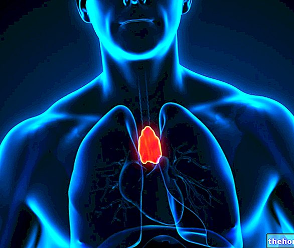
The rib cage is the skeletal structure that serves to protect vital organs (such as the heart and lungs) and important blood vessels (aorta, hollow veins, etc.).
Located in the upper part of the human body, exactly between the neck and diaphragm, the rib cage includes:
- Posteriorly, 12 vertebrae;
- Latero-anteriorly, 12 pairs of ribs (or ribs);
- Anteriorly, the costal cartilages and a bone called the sternum.
Each pair of ribs originates from one of the 12 posterior vertebrae, which are part of the rib cage.
In the anterior part, the ribs end with the costal cartilages; the latter represent the point of union with the sternum only for the first 7 pairs of upper ribs. In fact, from the eighth to the tenth pair, the single ribs join (again through cartilage) to the upper rib (therefore the octaves to the seventh, the ninths to the octaves etc); while from the tenth to the twelfth pair, they are free.
Between the ribs are numerous muscles, known as the intercostal muscles. The intercostal muscles allow the rib cage to expand during breathing; therefore they play a fundamental role in the introduction of air into the lungs.

In fact, in the first case, the coast is broken and often also in an unnatural position; a cracked rib, on the other hand, is "simply" bruised, therefore mostly intact and in the correct position.
particularly violent can lead to the breaking of the bones that make up the rib cage.
Two possible sporting activities that can induce a stressful rib fracture are golf and rowing.
RISK FACTORS
According to doctors, risk factors for a rib fracture include:
- Osteoporosis. Osteoporosis is a systemic disease of the skeleton, which causes severe weakening of the bones. This weakening arises from the reduction in bone mass, which, in turn, is a consequence of the deterioration of the microarchitecture of the bone tissue.
Therefore, people with osteoporosis are more prone to fractures, because they have more fragile bones than normal. - Participation in contact sports. Practicing sports in which physical contact is required is at high risk of fractures, not only in the lower or upper limbs, but also in the chest.
The athletes most at risk are rugby, soccer, American football, ice hockey and basketball players. - Neoplastic lesions of the ribs. A malignant tumor, originating in a rib, weakens the latter, making it more fragile and particularly susceptible to fractures.
EPIDEMIOLOGY
The ribs that most often suffer a fracture are those located in the center of the rib cage.
Fractures of the upper (first and second) ribs usually occur after facial trauma or blows to the head.
IF THE FRACTURE IS DUE TO TRAUMA
Often, when there is trauma at the "origin of the fracture", two signs appear on the "thoracic area involved in the" impact that certainly do not go unnoticed: swelling and hematoma.
IF THE FRACTURE IS MULTIPLE: POSSIBLE RISKS
If the rib fracture is multiple, it can lead to the onset of a potentially fatal medical condition, identified with the term "costal volet".
The costal volet consists of a partial or complete detachment of a group of ribs from the remaining rib cage. This can result in a paradoxical movement situation, in which the low-cut rib group makes movements opposite to those of the remaining rib cage.
The costal volet can be lethal when it causes a pneumothorax associated with severe respiratory failure. In fact, in such conditions, the lungs stiffen and breathing gradually becomes more and more difficult.
According to an Anglo-Saxon statistical study, for every 13 individuals who come to the hospital for a rib fracture there is one with a costal volet.
Some synonyms of costal volet are: mobile rib flap, mobile thoracic flap and flail chest.
WHEN TO SEE THE DOCTOR?
If they experience severe, permanent pain and have trouble breathing, people with severe chest trauma should see their doctor or go to the nearest hospital.
COMPLICATIONS
If severe or left untreated, a fracture of one or more ribs can lead to several complications, including:
- Major thoracic blood vessel injury. This occurs when the rupture affects the first three pairs of upper ribs. To cause damage to the aorta or to the other large vessels of the thorax is one of the two sharp bone stumps resulting from the fracture.
- Injury to one of the lungs. The ribs that, if fractured, can damage the lungs are those located in the middle of the rib cage. As before, the lungs are "stung" by one of the two pointed bone stumps, which are created after the broken bone breaks.
The main consequence of a rib affecting a lung is the collapse of the lung itself, due to the entry of air and blood into the pleural cavity. In medicine, this condition is also known as pneumothorax (PNX). - Injury to the spleen, liver or kidneys. These three organs are at risk of damage when the fracture affects the lower ribs and is such as to create very pointed extremities.
- Pneumonia and other lung disorders. The inability to breathe deeply, because this action causes pain, can lead to the onset of even severe lung inflammation.
Differences from the cracked rib
The symptomatic aspect that most differentiates a rib fracture from a crack is the fact that, in the second case, there is no risk of injury to the internal organs of the thorax.
, swelling, etc.), and asks him about the symptoms:
- What do they consist of?
- Following what event did they appear?
- What movements or gestures heighten its intensity?
Questions of this kind allow us to broadly understand the basic problem and what caused it.
After the questionnaire, the physical examination ends with the palpation of the painful area (to see what the patient's response is), the auscultation of the lungs and heart (in search of any abnormal sounds) and the analysis of the head, neck , spinal cord and belly.

INSTRUMENTAL EXAMINATIONS
Instrumental examinations are fundamental, as the information they provide allows the achievement of a correct and safe final diagnosis.
The prescribed procedures may include:
- X-rays. They allow to identify most of the rib fractures.
In fact, they present limitations only in the presence of "fresh" and not clear rib fractures.
X-rays are ionizing radiation that is harmful to health; however it should be remembered that the dose of such radiation is minimal. - CT scan. It provides a series of three-dimensional images, which very clearly reproduce the internal anatomy of the body.
It is very useful for analyzing not only the bones of the entire rib cage, but also the health of the thoracic blood vessels, lungs and abdominal organs.
It is based on the use of non negligible quantities of ionizing radiation. - Nuclear magnetic resonance (NMR). It is a radiological examination that provides for the patient to be exposed to completely harmless magnetic fields, without the need for harmful ionizing radiation.
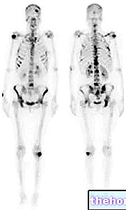
Like the CT scan, it is useful for evaluating a wide range of elements: ribs, blood vessels passing through the chest, lungs and organs of the abdomen. - Bone scan. It is a very sensitive nuclear medicine test, as it shows any bone alteration, even the least obvious.
Because of its sensitivity, doctors prescribe it when they suspect minimal fractures, barely visible through previous instrumental examinations. Such fractures are those that can cause a repetitive gesture or a strong cough.
Unfortunately, it is a somewhat invasive diagnostic technique. In fact, it involves the venous injection of a radioactive drug.
THE IMPORTANCE OF REDUCING PAIN
The planning of a drug treatment that reduces pain is of fundamental therapeutic importance. In fact, after the painful sensation has been reduced, the patient is able to take deep breaths again and this greatly reduces the risk of pneumonia.
PREVENT PNEUMONITIS
To prevent pneumonia from developing, doctors recommend coughing or taking a deep breath once or twice every hour.
CURIOSITY: THE THERAPY OF THE PAST
At one time, doctors treated rib fractures by applying a bandage to the patient's chest and trying to immobilize the affected area as much as possible. In other words, they acted almost like a fractured limb.
When they realized that this type of treatment, by limiting deep breathing, predisposed to pneumonia, they abandoned it, turning to the current method of treatment.

