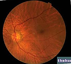
The macula is the central area of the retina, responsible for central vision; if its state of health is not optimal - as in the case of macular dystrophy - it is difficult to perceive colors, to read, to grasp the details of an object, to see clearly. clear and spotless etc.
Researchers agree that there are at least three variants of macular dystrophy: Stargardt's disease, vitelliform macular dystrophy and North Carolina macular dystrophy; all three are very rare morbid conditions.
The diagnostic process includes various procedures, including retinal fluorangiography, OCT and electroretinography.
Unfortunately, macular dystrophies are incurable pathologies, because they are supported by genetic mutations.
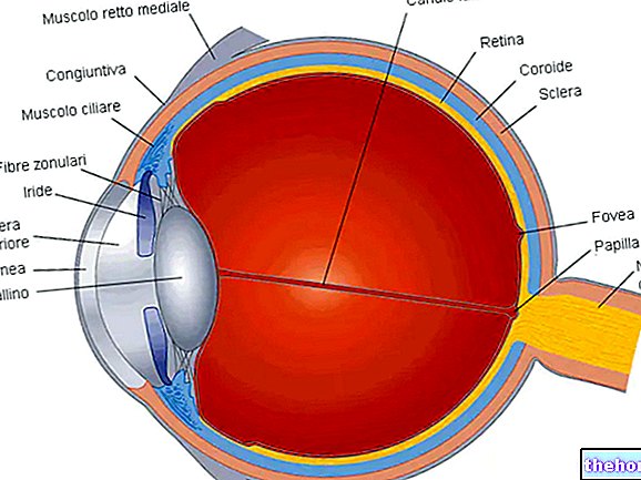
Brief review of the "Anatomy of the Eye"

In the eye (or eyeball), which takes place in the orbital cavity, three concentric portions can be recognized, which, from the outside towards the inside, are:
- The external cassock (or fibrous cassock). It is the area where the sclera (posteriorly) and cornea (anteriorly) reside; acts as an attachment for the so-called extrinsic muscles of the eyeball.
It possesses a fibrous nature. - The medium tunic (or uvea). It is a connective tissue membrane, rich in blood vessels and pigment.
Interposed between the sclera and the retina, it is responsible for providing nourishment to the retina, or rather to the layers of the retina with which it comes into contact.
It includes iris, ciliary body and choroid. - The inner cassock. It basically consists of the retina. The latter is a transparent film made up of ten layers of nerve cells (or neurons), including the so-called cones and rods responsible for visual function.
In light of this, the inner tunic has a nervous function.
What does "dystrophy" mean?
In medicine, the term "dystrophy" refers to the structural and functional degeneration of an organ or tissue, following a process of cellular involution (atrophy).
What is the Macula?
The macula (or macula lutea) looks like a yellow spot of about 5.5 millimeters in diameter.
Located to the side of the optic nerve emergence at a distance of about 2.5 centimeters, it represents the retinal area with the greatest visual acuity and the greatest ability to identify details.
Furthermore, as it contains more cones than rods, it is particularly sensitive to light stimuli and the perception of colors.
All the aforementioned characteristics describe the so-called central vision.
At least four regions can be recognized in the macula: two of these are particularly important and are identified with the names of fovea and foveola (N.B: the foveola is the center of the fovea).
As far as vascularization is concerned, the fovea has large blood vessels all around it and arterioles, venules and capillaries inside it; the foveola, on the other hand, is avascularized.
The yellow color that distinguishes the macula is due to the presence of two carotenoids: lutein and zeaxanthin.
Macular Dystrophy and Macular Degeneration: The Link
Macular dystrophy can be considered an early-onset (or juvenile) form of macular degeneration.
In medicine, the term "macular degeneration" refers to a group of eye diseases characterized by damage to the macula and progressive loss of vision.
The most well-known and widespread form of macular degeneration - which despite the similar symptoms should not be confused with macular dystrophy - is the so-called senile macular degeneration (linked to advanced age). This morbid condition is typical of elderly people and can be attributed to the natural aging process to which every human being is subjected.
For further information: Macular Degeneration: What is it? DNA.According to what emerged from the most recent scientific researches, in most cases these mutations would be transmitted by inheritance (ie from one of the two parents); however, it is good to remember that, in some cases, they appear unexpectedly and without an explainable reason in the course of life.
Types of Macular Dystrophy
Researchers have identified at least three different types of macular dystrophy:
- Stargardt's disease. It is the most common macular dystrophy of those known.
There are two subtypes, one affecting children / adolescents between the ages of 6 and 13 and one affecting young adults aged 18-20.
According to the latest discoveries, there are more than one genes that, if mutated, can cause this ocular pathology; the best known and most studied is certainly ABCA4.
A fairly typical feature of Stargardt's disease is the presence of deposits of lipofuscin, a toxic pigmented substance, inside the cells of the retina (and therefore also of the macula).
Generally, it is an autosomal recessive inheritance disorder. - Vitelliform macular dystrophy. Those who suffer from this type of macular dystrophy present, in the fovea of the macula, a yellow lesion of moderate size, very similar in shape to the yolk sac (hence the term vitelliform).
As with Stargardt's disease, there are two variants of vitelliform macular dystrophy: a variant that affects children and is a hereditary disease (Best's disease) and a variant that affects young adults and derives from a genetic mutation acquired in the course of life.
The researchers identified at least two genes responsible for the disease: BEST1, which is linked to Best's disease, and PRPH2, which is associated with the young adult-onset variant.
Furthermore, they found, even in this circumstance, the frequent presence of lipofuscin deposits in the cells of the macula.
As a hereditary disorder, vitelliform macular dystrophy is an autosomal dominant inheritance disorder. - Macular dystrophy of North Carolina. Information relating to this eye disease is scarce. It is only known that it is an autosomal dominant hereditary disease with juvenile onset that causes the "accumulation of" drusen "in the cells of the retina," drusen "are small protein-lipid deposits.
The reference to North Carolina is explained by the fact that the first clinical cases were identified in this US state.
Epidemiology: How Widespread is Macular Dystrophy?
Macular dystrophy is a very rare condition.
While the prevalence rate (one case per 8,000-10,000 births) has been calculated for Stargardt's disease, no epidemiological data has yet been established with certainty for vitelliform macular dystrophy and North Carolina macular dystrophy.
:
- Wavy and / or blurry vision. A typical disturbance is seeing straight lines as wavy or arched;
- Impaired vision of colors. As you will remember, the macula is the area of the retina used for the perception of colors;
- Loss of vision and reduction of visual acuity;
- Difficulty adjusting vision in poorly lit places;
- Vision of blind spots;
- Problems in identifying the details of what is being observed;
- Vision of one or more black spots in the center of the visual field;
- Difficulty in reading, especially if the characters are very small, and in driving.
When macular dystrophy is in its infancy, the symptoms are mild. For example, at this stage, patients may not perceive some details of an object or person.
As the disease progresses, the disorders become more and more marked and incompatible with a normal life. It is typical of this moment to see black spots instead of a person's face, while looking straight in the eye.
Macular Dystrophy: Why Do You Lose Your Vision?
The loss of vision, which occurs in the presence of macular dystrophy, is due to the progressive death of the retinal photoreceptors and the involvement of the retinal pigment epithelium.
Macular Dystrophy and Peripheral Vision
Since the deterioration is limited to the macula, peripheral vision is completely normal in people with macular dystrophy.
Therefore, when a patient looks a person in the face, he claims to clearly distinguish his arms, legs, etc.
Macular Dystrophy: Complications
Since the genetic mutations underlying macular dystrophy cannot be healed, vision problems are irreversible and tend to slowly worsen as time passes.
A possible complication of Best's disease is the macular hole.
When to contact the doctor?
If you experience one or more of the aforementioned symptoms and if you belong to a family with a history of macular dystrophy, you should first contact your doctor and then an ophthalmologist, i.e. a doctor who specializes in eye diseases.
The in-depth study of the current situation allows us to know if it is a form of progressive macular dystrophy or not.
Fundus examination
The examination of the ocular fundus allows to analyze the internal structures of the eyeball, therefore also the retina and the macula.
It can provide various interesting indications, even if often, for a better final evaluation, it is necessary to resort to more specific examinations.
Although it requires the use of eye drops to dilate the eye pupil, it is not a particularly invasive test.
Computed Optical Tomography (OCT)
Computed optical tomography (OCT) is a reliable and non-invasive diagnostic test that provides very precise scans of the cornea, retina, macula and optic nerve.
With a total duration of 10-15 minutes, it is performed using an instrument that emits a laser beam free of harmful radiation. Obviously, the patient should be made to sit in front of this instrument, so that the laser penetrates the patient's eyes.
The latest generation OCTs work well even without having to administer eye drops to the individual under examination to dilate the pupils.
In the case of macular dystrophy, the scans produced by the diagnostic tool are able to show the deposits of lipofuscin at the level of the retinal pigment epithelium.
The lipofuscin deposits - which are essentially made up of waste products - appear as yellow-brown presences.
Retinal Fluorangiography
Retinal fluorangiography (retinal fluorescence angiography) is a photographic diagnostic procedure, which allows the identification and study of vascular pathologies of the eye.
The examination involves the venous injection of a dye, called fluorescein, and the observation of its diffusion in the bloodstream using a special instrument, called a retinograph.
The retinograph is able to take real photographs (or frames) of the blood flow inside the vessels of the retina.
The examination lasts about 10 minutes and, at the time of the dye administration, it could be slightly annoying.
In the presence of macular dystrophy, the frames are able to identify the areas of the macula that have suffered deterioration.
Electroretinography (ERG)
Electroretinography (ERG) is a diagnostic test that allows to measure the electrical response of the retina - for the accuracy of the photoreceptors of the retina - to one or more light stimuli.
As for its execution, corneal and skin electrodes are needed, which record the electrical signal coming from the retina, and an instrument that emits light beams of different intensity.
Before the procedure, obviously, eye drops for pupil dilation should be instilled in the patient's eye.
ERG is particularly suitable for all hereditary macular degenerations, therefore not only for macular dystrophy but also for retinopathy pigmentosa (which is another form of hereditary macular degeneration).
It can also be very useful when these pathologies are not yet particularly evident from the symptomatological point of view.
Macular Dystrophy and Aids for the Visually Impaired
For patients with severe vision problems, we remember the existence of aids for the visually impaired, which, nowadays, are of enormous help in various daily activities.


