Generality
Macular degeneration is a condition in which the central portion of the retina (called the macula) deteriorates and does not function properly. The disease is often referred to as age-related macular degeneration (AMD or AMD), as it occurs mainly in people over the age of 60. Many elderly people, in fact, develop the disease as part of the natural aging process.
Some cases of macular degeneration are mild and do not completely affect vision, while other forms are severe and can cause vision loss in both eyes.
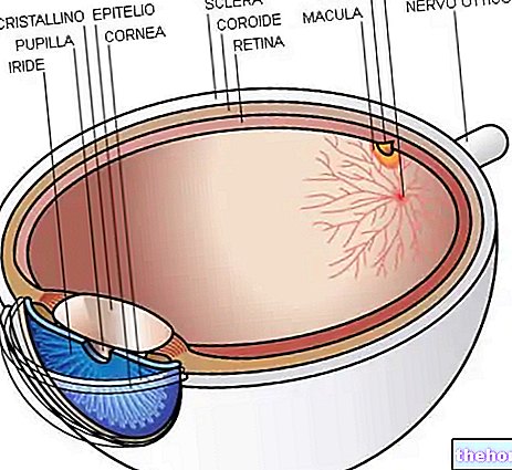
Note. Macular degeneration affects the macula, a small central portion of the retina (a layer of photosensitive tissue that lines the back of the eye).

Types of macular degeneration
Two main forms of age-related macular degeneration can be distinguished: dry and wet.
Dry macular degeneration occurs when small yellowish protein and glycemic deposits, called "drusen", begin to accumulate under the retina, due to blood reabsorption. Due to the presence of drusen, the macula can become thinner and stop functioning properly, leading to gradual darkening of vision. In the more advanced stages of the disease, the thinning of the layer of photosensitive cells can lead to tissue atrophy or death. Also, in some cases, dry macular degeneration can progress to the wet form.
Wet (or wet) macular degeneration accounts for only 10% of all cases. The disease is characterized by the growth of abnormal blood vessels from the choroid, at the macula (choroidal neovascularization). The deformation and distortion of vision is caused by the leakage of blood and fluids from the newly formed blood vessels, which collect under the macula and lift it. Wet macular degeneration is more aggressive than the dry form, as it can cause rapid and severe loss of central vision (caused by scarring of the blood vessels).
Juvenile macular degeneration
Different forms of macular degeneration affect children, teens or adults. Many of these juvenile (or early onset) diseases are inherited and are more correctly referred to as macular dystrophies.

Stargardt's disease is the most common form of juvenile macular dystrophy. The condition typically develops during childhood and adolescence and is almost always inherited as an autosomal recessive trait (ie it occurs only when a child inherits two copies of the altered ABCA4 gene, each from parents who carry the disease). The hallmark of Stargardt's disease is decreased central vision. The progressive loss of vision, associated with the disease, is caused by the death of photoreceptor cells in the macula and by the involvement of the retinal pigment epithelium.
Symptoms
For further information: Senile Macular Degeneration Symptoms
Macular degeneration is usually bilateral, although the clinical appearance and degree of visual loss can vary greatly between the two eyes; if only one eye is involved, the changes in vision may not be noticeable because the other will tend to compensate for low vision.
- Symptoms of dry macular degeneration include blurred central vision or the presence of a small blind spot in the visual field. Over time, the blind spot becomes progressively larger and further impairs vision, making reading, driving, or other daily activities more difficult.
- Symptoms of wet macular degeneration usually onset and worsen rapidly, leading to sudden loss of central vision. Manifestations of the disease include distorted, confused, or irregular vision.
Regardless of the type of macular degeneration, the most common symptoms include:
- Decreased visual acuity;
- Difficulty seeing in bright environments (photophobia);
- Need for an increasingly brighter light source to see closely;
- Difficulty or inability to recognize people's faces
- Difficulty adapting from dark to light.
Macular degeneration almost never causes complete blindness, as it does not affect peripheral vision (it does not affect the entire retina), but it can cause significant visual impairment. For example, with advanced macular degeneration, one can distinguish the outline of a clock, but the patient may not be able to see the clock hands to tell what time it is.
Causes and Risk Factors
The exact cause of macular degeneration still remains unknown. However, many experts believe that some risk factors contribute to the development of macular degeneration.
The biggest risk factor is age. Studies show that people over 60 are clearly more at risk: by the age of 65, the macula begins to degenerate in about 10% of patients. The prevalence of damage increases to 30% in subjects aged 75-85.
Inheritance is another risk factor for macular degeneration. People who have a close relative who has the disease are more likely to develop macular degeneration.
Other risk factors include smoking, obesity, Caucasian population, female sex, a diet low in fruit and vegetables, prolonged exposure to sunlight or other types of ultraviolet light, hypertension and high levels of blood cholesterol.
Diagnosis
Many people are unaware that they have macular degeneration until they have significant vision problems or until the condition is identified during an eye examination. Early diagnosis of age-related macular degeneration is very important, as some treatments are available that can delay or reduce the severity of the disease.
For the diagnosis of dry macular degeneration, a complete examination of the eye with an ophthalmoscope, a device that allows you to see the retina and other structures of the back of the eye, may be sufficient. If the ophthalmologist suspects the wet form, fluorangiography and optical coherence tomography (OCT) may be done.

Optical coherence tomography (OCT) can accurately highlight areas where the retina is thinnest or where edema is present.
A visual acuity test helps determine the extent of central low vision. The Amsler Grid Test, one of the simplest and most effective methods of monitoring macular health, can be used to detect both types of macular degeneration. Amsler's grid is, in essence, a pattern of intersecting straight lines (similar to graph paper), with a black dot in the middle.In this test, the patient covers one eye and stares at the central black point, keeping the grid 12 to 15 centimeters away from the face. With normal vision, all grid lines surrounding the black point are straight, evenly spaced, with no missing or abnormal-looking areas. If looking directly at the center point with the eye uncovered, the lines surrounding it appear bent, distorted and / or missing, a disease affecting the macula may be suspected.
People who develop macular degeneration should have regular examinations to constantly monitor the progression of the disease and, if necessary, initiate treatment.

