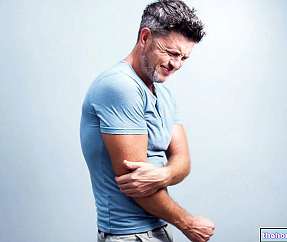
The main causes of elbow bursitis are: trauma to the elbow, prolonged pressure in the elbow, some forms of arthritis and infections following cuts, wounds or insect bites in the elbow.
Typical symptoms and signs of an elbow bursitis consist of: swelling, pain and difficulty moving.
Typically, diagnosis is based on physical examination and history; rarely, doctors resort to more in-depth examinations.
First-line treatment is conservative. The use of more invasive treatments (surgery) occurs only when conservative therapies have not provided the desired results.

Brief reminder of the anatomy of the elbow
The elbow joint results from the relationship of three bony elements: humerus, ulna and radius.
The humerus is the bone of the arm; ulna and radius, on the other hand, are the bones of the forearm.
In its distal end, the humerus has two characteristic areas: the so-called capitulum and the so-called humeral trochlea.
- THE capitulum it is the part of the humerus that articulates with the head of the radius. The head of the radius represents the most proximal end of this bone;
- The humeral trochlea is the part of the humerus that articulates with a particular portion of the ulna, called the olecranon. Much like a hook, the olecranon represents the most proximal end of the ulna.
The synovial bags represent an extremely important part of the anatomical elements characteristic of the synovial joints.
A generic synovial bursa is a sac, lined with a synovial membrane and containing a fluid, known as synovial fluid.
Acting as anti-friction and anti-friction pads, the synovial bursae have the task of preserving the ligaments, tendons, cartilage tissues and other anatomical structures of the synovial joints.

Elbow bursitis due to infections has characteristic clinical signs.
Failure to treat an infectious elbow bursitis can lead to the spread of bacteria into the bloodstream, which have infected the synovial bursa near the olecranon.
The spread of these bacteria in the bloodstream can cause unpleasant consequences and endanger the life of those who suffer from it. it occurs only in the case of suspected presence of an osteophyte at the level of the olecranon of the ulna. Osteophytes are small bone spurs, similar to a rose thorn, a beak or a claw, which form along the joint margins of bones subjected to chronic erosive and irritative processes.
The sampling and analysis of the fluid present in the swollen synovial bursa takes place only if there is a possibility that the bursitis is due to an infection or episode of gout.
Identifying the precise causes of an elbow bursitis is essential for planning the most appropriate therapy.
it is essential to fight infecting bacteria and prevents unpleasant consequences (eg: spread of bacteria in the blood).SURGICAL THERAPY
Surgical therapy, foreseen in case of elbow bursitis, consists in the removal of the inflamed synovial bursa.
If the bursitis had an infectious origin, post-operative antibiotic treatment and at least one day of hospitalization are foreseen.
If, on the other hand, the bursitis had a "non-infectious origin, the patient does not need anything else" and can return home on the day of the operation, usually after a few hours from the end of the procedure.
Generally, the recovery phase from elbow bursitis surgery does not require special physiotherapy (but only some exercises that the patient can do at home) and has a canonical duration of 3-4 weeks.
Does the synovial bursa reform?
Once eliminated by surgery, the synovial bags have a tendency to reform in their original location.
Reformation of a synovial bursa is a very slow process, which can take many months.


