Generality
The ureter is an even, symmetrical tubular canal that connects each kidney to the bladder.
About 28-30 centimeters long and having an average diameter of about 6-8 millimeters, it has three portions: abdominal, pelvic and bladder.

Figure: anatomy of the kidney. Thanks to the image, the reader can appreciate the precise location of the renal pelvis, the major calyxes and all those other structures mentioned in the article.
The abdominal portion constitutes the first section of the ureteral canal, after its birth at the level of the renal pelvis.
The pelvic portion represents the second section, the one with origin at the level of the pelvic cavity and ending at an antero-medial curvature of the ureteral canal.
The bladder portion, finally, is the last section, opening, with the ureteral orifice, inside the bladder.
The function of the ureters is to carry urine, produced by the kidneys, into the bladder.
Brief anatomical recall of the urinary system
The elements that make up the urinary tract are the kidneys and the urinary tract.
The kidneys are the main organs of the excretory system. Two in number, they reside in the abdominal cavity, on the sides of the last thoracic vertebrae and the first lumbar vertebrae, they are symmetrical and have a shape that resembles that of a bean.
The urinary tract, on the other hand, forms the so-called urinary tract and presents, from top to bottom, the following structures:
- The ureters, the description of which belongs to this article.
- The bladder. It is a small hollow muscular organ, which accumulates urine before urination. It resides in the pelvic cavity.
- The urethra. It is the tubular shaped canal that connects the bladder to the so-called urinary meatus (or external urethral orifice) and which is mainly used for the expulsion of urine.

So is the ureter
The ureter is an even duct, symmetrical and of moderate diameter, which connects each kidney to the bladder and carries the urine to be expelled inside the latter.
In other words, the ureter is a drainage tube, which favors the advancement of urine towards the structures responsible for the urination process.
Obviously, from the right kidney, the so-called right ureter originates and, from the left kidney, the so-called left ureter.
Anatomy
With origin from the renal pelvis (abdomen) and termination at the level of the bladder (pelvic cavity), the ureter has an average length of about 28-30 centimeters and an average diameter of about 6-8 millimeters (NB: the diameter varies considerably to depending on the point considered).
Anatomical experts recognize three portions in the ureter: the abdominal portion, the pelvic portion and the bladder portion.
In addition to the course of the ureters, in this chapter, the reader will be able to find information regarding their anatomical relationships, their histological structure, their blood supply and their innervation.
Brief review of the concepts: sagittal plane, medial position and lateral position
In anatomy, medial and lateral are two terms with opposite meanings. However, to fully understand what they mean, it is necessary to step back and review the concept of the sagittal plane.

Figure: the planes with which anatomists dissect the human body. In the image, in particular, the sagittal plane is highlighted.
The sagittal plane, or median plane of symmetry, is the antero-posterior division of the body, from which two equal and symmetrical halves derive: the right half and the left half. For example, from a sagittal plane of the head derive a half, which includes the right eye, the right ear, the right nasal nostril and so on, and a half, which includes the left eye, the left ear, the left nasal nostril etc.
Returning therefore to the medial-lateral concepts, the word medial indicates a relationship of proximity to the sagittal plane; while the word lateral indicates a relationship of distance from the sagittal plane.
All anatomical organs can be medial or lateral to a reference point. A couple of examples clarify this statement:
First example. If the reference point is the eye, this is lateral to the nasal nostril on the same side, but medial to the ear.
Second example. If the reference point is the second toe, this element is lateral to the first toe (big toe), but medial to all the others.
ABDOMINAL PORTION
So called because it takes place at the level of the abdomen, the abdominal portion of a ureter is its initial (or proximal) section
The starting point coincides with the so-called renal pelvis (or renal pelvis). Located within the renal hilum, the renal pelvis is that area of each kidney that receives urine from the major calyxes. In fact, it marks the passage between the kidneys and the urinary tract.
At where the ureter arises, the renal pelvis narrows, giving rise to the so-called uretero-pelvic junction.
From the uretero-pelvic junction, the ureter follows a downward path, which leads it to pass anteriorly to the great psoas muscle and always remain in the retroperitoneal position, until it enters the pelvis.
As it enters the pelvis (the area after which the pelvic portion begins), the ureter passes close to the common iliac arteries.
Reports of the abdominal portion
The abdominal portion of the two ureters borders from top to bottom:
- Sideways (i.e. on the outer side), with the lower pole of the kidney, the ascending colon (right ureter) and the descending colon (left ureter).
- Dorsally, with the great psoas muscle, the genitofemoral nerve and the common iliac arteries.
- Medially (ie on the internal side), with the inferior vena cava (right ureter), the internal spermatic vein (left ureter), the so-called orthosympathetic chain and the lumbar lymph nodes.
- Anteriorly, with the parietal peritoneum of the posterior wall of the abdomen, the spermatic vessels (only in the male) and the ovarian vessels (only in the female).
PELVIC PORTION
The pelvic portion of each ureter is the section that takes place in the pelvic cavity.
First, it runs along the lateral pelvic walls; later, in this case at the level of the ischial spines, it undergoes a curvature in an antero-medial direction, which leads the ureteral duct to assume a slightly transverse position with respect to the bladder.
The antero-medial curvature of the ureters is essential to avoid the reflux of urine, from the bladder towards the kidneys.
Relationships of the pelvic portion
In the two sexes, the pelvic portion of the two ureters establishes slightly different relationships, as the pelvic anatomy of men and women is different.
- Later, adjoins the hypogastric vessels (in both men and women).
- Medially, is in relation, from top to bottom, with the rectum (both sexes), the pelvic fascia covering the levator muscle of the anus (only in men), the vas deferens (only in men), the lateral margin of the bladder (only in men), the seminal vesicle (only in men), the ovarian dimple (only in women), the infundibulum of the uterine tube (only in women), the uterine artery (only in women) and the wall of the bottom of the bladder (only in women).
VESCICAL PORTION
The bladder portion of each ureter is the section that communicates with the bladder.
10-15 millimeters long, it crosses the bladder wall obliquely until it reaches the bladder cavity. Here, it forms an opening which takes the name of ureteral orifice.
The oblique crossing of the bladder wall is the result of the antero-medial curvature, which the pelvic portion of each ureter undergoes.
Maintaining the oblique disposition helps to prevent the reflux of urine from the bladder towards the kidneys.
TONACHE AND EPITHELES OF THE "URETER: A LITTLE" OF HISTOLOGY
The wall of each ureter has three cassocks, which, from the inside outwards, are: the mucous cassock, the fibromuscular cassock and the adventitious cassock.

Figure: thanks to the image, the reader can appreciate the antero-medial curvature of the ureters, at the level of their pelvic portion.
Without going into too much detail, the mucous membrane has mainly transitional epithelium, an elastic cell lining typical of the urinary tract (so much that experts also call it urothelium).
The fibromuscular cassock contains mostly smooth muscle cells, interspersed with bundles of connective tissue.
Finally, the adventitious cassock comprises loose connective tissue, characterized by elastic fibers. Its presence is considerable at the level of the bladder portion.
BLOOD SPRAYING OF THE URETERS
The arterial vessels of each ureter arise from the renal, genital and hypogastric arteries.
In the case:
- The renal artery deals with the arterial supply of the upper tract of each ureter.
- The "genital artery" affects the arterial supply of the median tract of each ureter. Derivation of the abdominal aorta, the genital artery takes the specific name of testicular artery in men and ovarian artery in women.
- The hypogastric artery deals with the arterial supply of the lower tract of each ureter. Also known as the internal iliac artery, the hypogastric artery has numerous branches, all of which participate in the blood supply of the ureters.
Table. The branches of the hypogastric artery, which participate in the blood supply of the lower part of the ureters.
- The superior bladder artery
- The uterine artery (only in women)
- The middle rectal artery
- Vaginal arteries (only in women)
- The inferior bladder artery (only in humans)
As far as the venous vessels are concerned, these flow from top to bottom:
- In the venous network of the adipose capsule of the kidney
- In the renal vein
- In the spermatic venous plexus (only in men) and in the ovarian venous plexus (only in women)
- In the branches of the hypogastric vein
INNERVATION OF THE URETERS
The nerves that innervate each ureter are sympathetic and parasympathetic nerve fibers, which arise from the renal, testicular (in men) / ovarian (in women) and bladder plexuses.
The sympathetic fibers belong to the sympathetic nervous system and have an inhibitory action against urination; the parasympathetic fibers, on the other hand, belong to the parasympathetic nervous system and promote urination.
Insight into the sympathetic nervous system and parasympathetic nervous system
Together, the sympathetic nervous system and the parasympathetic nervous system constitute the so-called vegetative (or autonomous) nervous system, which performs a fundamental control action of involuntary bodily functions.
The sympathetic nervous system tends to be active during an emergency situation. Not surprisingly, doctors claim that he presides over the "fight and flight" adaptation system.
On the other hand, the sympathetic nervous system tends to be activated in situations of stillness, rest, relaxation and digestion. For this reason, doctors consider it to be the basis of the "rest and digestion" adaptation system.
* Please note: in the medical field, the word "plexus" is used both when talking about blood vessels and when talking about nerves. A vascular plexus is distinctly different from a nerve plexus: the former is a reticular formation of intertwined arterial (or venous) vessels, while the latter is a reticular formation of nerves.
Functions
Each ureter has the important function of conducting urine from the kidneys into the bladder.
Diseases of the Ureter
Among the problems that can affect the ureters, one of the most relevant and widespread is the so-called ureteral stones.
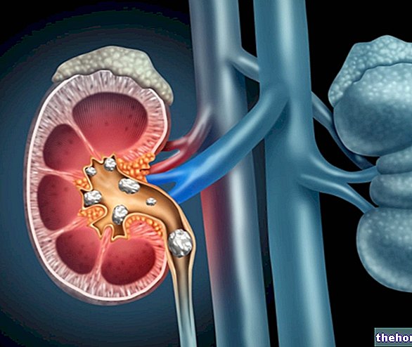
Similar to kidney and bladder stones, ureteral stones is a pathological condition of the urinary tract, characterized by the presence of small mineral aggregates within one or both ureters. These mineral aggregates (commonly called calculi) derive from the precipitation of certain substances contained in the urine and, as a result of their accumulation, can obstruct the ureters that contain them.

Figure: ureteral stones, kidney stones and bladder stones.
With obstruction of one or both ureters, urine flow is inadequate and symptoms such as pain when urinating and / or hematuria (blood in the urine) appear.
Inside the ureteral duct, there are sections that are most affected by ureteral stones, due to their particular narrowness (in diameter). These sections are: the uretero-pelvic junction, the last section of the abdominal portion and the portion of ureter that joins the bladder (bladder portion).





.jpg)


