Generality
Cardiac arrhythmias are changes in the heart's normal rhythm of contraction. These anomalies, it will be seen, do not concern only the number of heartbeats per minute, but also the propagation of the impulse that generates them.

The therapy to be adopted depends on the cause that determines the arrhythmia. Despite this, there are basic therapeutic interventions, valid in any episode of arrhythmia; the generic treatment consists in the administration of antiarrhythmic drugs and beta-blockers, in the use of particular medical instruments and in adopting healthy lifestyles, if the individual with an arrhythmia is used to smoking or drinking excessively.
The heart
To fully understand what an arrhythmia is and what triggers it, it is good to remember some characteristics of the heart regarding its ability to self-control.
The myocardium, that is the muscular tissue of the heart, has some cells that are distinguished, compared to all the other cells of the human body, for two unique properties: the automaticity and rhythm of the nervous impulse intended for contraction. By automaticity we mean the ability to spontaneously and involuntarily initiate the contraction activity of the myocardial cells, generating the nerve impulse by itself. This is a real exception, as the other muscle cells in the body work differently: for example, if you want to bend an arm to lift a weight, the signal starts from the brain and reaches the muscles of the limb. In the heart, instead , the signal originates from the muscle cells themselves and is not controlled by a central system such as the brain.
The second exclusive property is the rhythmicity of the spontaneous activity of contraction. It consists in the regularity and in the ordered succession in time of the nervous impulse.
Therefore:
- Automaticity: it is the ability to form impulses of muscle contraction spontaneously and involuntarily, that is, without an input coming from the brain.
- Rhythmicity: it is the ability to neatly transmit the impulses of muscle contraction.

- Sino atrial node →
- Atrioventricular node →
- Bundle of His (Atrioventricular bundle) →
- Purkinje fibers.
- Like all other muscle cells, these too, after the passage of the contraction impulse, are insensitive to another impulse very close in time. In other words, after a first impulse, the myocardial cells need time to respond to a subsequent impulse This amount of time, necessary for muscle cells to restore receptivity, is called refractoriness.
It will be seen that a change in the dominant center and refractoriness can have repercussions on the regularity of the heartbeat.
Finally, the last piece of information not to be forgotten concerns the cardiac cycle. The cardiac cycle is the alternation of a phase of myocardial contraction, called systole, and a relaxation phase, called diastole. During the contraction, blood is pumped through the efferent vessels; conversely, the relaxation of the myocardium allows blood to flow into the heart through the afferent vessels.
What are cardiac arrhythmias and how are they classified
Cardiac arrhythmias are changes in the normal heartbeat rhythm. There are three possible alterations and it is sufficient that one is present for an "arrhythmia to arise. They are:
- Changes in the frequency and regularity of the sinus rhythm.
- The variation of the seat of the dominant marker center.
- Impulse propagation (or conduction) disturbances.
1. Changes in the frequency and regularity of the sinus rhythm, ie the normal rhythm imposed by the sinoatrial node, result in the so-called tachycardias and bradycardias. Tachycardia is an increase in the heart rate, meaning the heart beats faster than normal. Conversely, bradycardia is a slowing of the heart rate, so the heart beats more slowly. There are two threshold values, expressed in beats per minute, which delimit the normal range: 60 beats per minute is the minimum value; 100 beats per minute is the maximum value. Below 60 beats, there is bradycardia; above 100 heartbeats, you have tachycardia.
The so-called physiological sinus arrhythmias also manifest frequency alterations. They are not alarming episodes, they occur more often at a young age and their causes are related to central metabolism and respiratory reflexes.
2. The variation of the seat of the dominant step center occurs when the sinoatrial node decreases or even loses its automaticity. This therefore determines its replacement with a secondary pathway center, such as the atrioventricular node. If the phenomenon is limited to a few cycles, we speak of extrasystoles, that is, premature beats; if the phenomenon persists for a succession of cycles, one encounters junctional and ventricular tachycardias and atrial and ventricular fibrillations. These are abnormal situations that should not be underestimated, since these alterations almost always manifest themselves in pathological circumstances.
3. Disturbances in the propagation (or conduction) of the impulse occur as a consequence of a slowing down, or arrest, of the impulse itself during the journey from the dominant pathway center to the secondary centers. The obstacle can be caused by an anatomical interruption of the conduction pathway, or by a difficult restoration of the faculty of response to an impulse (prolonged refractoriness). Refractoriness can be prolonged due to:
- Medicines.
- Neurogenic stimuli.
- Pathological conditions.
Once the alterations have been clarified, arrhythmias can be classified in at least two ways: on the basis of the physiopathological characteristics of the alterations and on the basis of the site of origin of the disorder.
The pathophysiology (i.e. the study of the functions changed due to a pathological condition) of the three alterations described above allows us to distinguish arrhythmias into two large groups:
- Arrhythmias mainly due to a modification of automaticity (or impulse formation). Arrhythmias with:
- Changes in the frequency and regularity of the sinus rhythm.
- Variation of the seat of the dominant marker center.
- Arrhythmias mainly due to a modification of the conduction (or propagation) of the impulse. Arrhythmias with:
- Impulse propagation disorders.
It should be noted that the difference between these two groups of arrhythmias is subtle. Very often, in fact, an arrhythmia due to a change in conduction can turn into one due to changes in automaticity. For example, when an obstacle downstream opposes the conduction of the impulse coming from the sinus node, this block causes the dominant marker center to change; the new dominant center, at that point, takes command of the rhythm. the opposite case is also true, that is, that arrhythmias due to modifications of automaticity change into arrhythmias caused by a modification of conduction; it is the case in which a high frequency increase does not leave the myocardial cells time to become receptive, consequently altering the propagation of the impulse.
The classification based on the site of origin of the disorder distinguishes arrhythmias in:
- Sinus Arrhythmias. The disorder concerns the impulse coming from the sinoatrial node. Generally, the frequency changes are gradual. Some examples:
- sinus tachycardia
- sinus bradycardia
- sinoatrial block
- Ectopic Arrhythmias. The disorder involves a pathway other than the sinoatrial node. Typically, they arise abruptly. The affected areas divide ectopic arrhythmias into:
- Supraventricular. The disorder affects the atrial area. Some examples:
- atrial flutter
- atrial fibrillation
- Atrioventricular, or nodal. The affected area concerns the atrioventricular node. Some examples:
- paroxysmal supraventricular tachycardia
- junctional extrasystole
- Ventricular. The disorder is located in the ventricular area. Some examples:
- ventricular tachycardia
- ventricular flutter
- ventricular fibrillation
- Supraventricular. The disorder affects the atrial area. Some examples:
It is common to use this second classification, but it should not be forgotten that it is closely linked to the first, since the change in the site of origin of the disorder is a direct consequence of one of the pathophysiological mechanisms described above.
Possible causes
Various causes contribute to determining the changes in automaticity and rhythm:
- Congenital heart disease, i.e. present from birth.
- Acquired heart disease, that is, developed over the course of life.
- Hypertension.
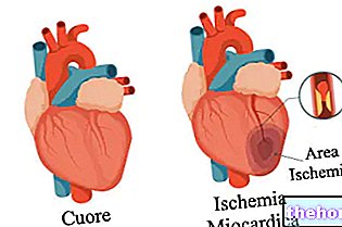
- Cardiac ischemia.
- Myocardial infarction.
- Hyperthyroidism.
- Alcohol and drug abuse.
- Smoke.
- Drug poisoning.
Acquired heart disease can arise regardless of a lifestyle characterized by alcohol and drug abuse. This is why both appear on the list. The same applies to the use of drugs.
More frequent symptoms
Symptoms are variable and would require a much longer description than the following. In fact, as we have seen, there are many arrhythmias, each with its own particular pathophysiology and caused by different factors. This means that the symptoms are numerous and the presence / absence of one of these distinguishes the single arrhythmia. In general, the symptomatological picture worsens in tandem with the severity of the arrhythmia manifested by a patient.
A list of the main symptoms is as follows:
- Tachycardia (or heartbeat / palpitation).
- Bradycardia.
- Irregular heartbeat.
- Dyspnea.
- Chest pain.
- Anxiety.
- Dizziness and vertigo.
- Sense of weakness.
- Fatigue after minimal effort.
It should be remembered that a heart rhythm that, in terms of beats per minute, remains within the 60-100 range is considered normal.
Diagnosis
A cardiological visit is the first step in diagnosing an "arrhythmia. It is based on:
- Pulse measurement.
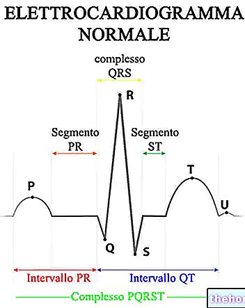
- Electrocardiogram (ECG).
- Dynamic electrocardiogram according to Holter.
Pulse measurement. It is a simple investigation, which can be done by anyone, not just the doctor. It does not have the same reliability as an instrumental examination, clearly, and does not inform about the characteristics of the arrhythmia.
Electrocardiogram (ECG). By measuring the electrical activity of the heart, ie that which allows the contraction of the myocardium, the ECG shows the great variety of arrhythmias that can occur in a patient. The different types of arrhythmias show different patterns from each other and the cardiologist, based on these results, can define the heart problem.
Dynamic electrocardiogram according to Holter. This diagnostic method works like a normal ECG, with the difference that the patient is monitored for 24-48 hours, without interruption. During this time, the patient is free to carry out normal activities of daily life. This investigation is required when the arrhythmia occurs sporadically. In fact, certain arrhythmias may occur as isolated episodes.
Therapy
As for the symptoms, the therapy to be adopted also depends on the type of arrhythmia and any associated heart disease. Therefore, the main therapeutic interventions, both pharmacological and instrumental, will be reported below.
The drugs administered are:
- Beta-blockers and calcium channel blockers. They are used to slow down the heart rate.
- Antiarrhythmics. They serve to stabilize the heart rhythm.
- Anticoagulants. They are used to thin the blood and are used to prevent the formation of thrombi or emboli in cases of particular arrhythmias, such as atrial fibrillation.
The main instrumental / surgical interventions are:
- Electrical cardioversion. It consists in "applying a single electric discharge, also called shock, to reset and restore the sinus rhythm, that is, the one marked by the sinoatrial node (dominant path marker center).
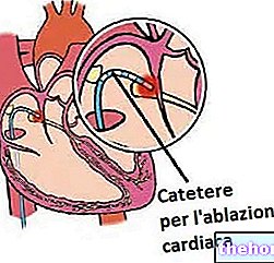
- Radiofrequency ablation, or catheter ablation. It is used in patients with tachycardias. It involves the use of a particular catheter, which is inserted into the femoral veins and brought to the heart. Through the catheter, two operations are carried out: first, an electrical discharge is infused into the heart to determine which area of the myocardium works in Once this is done, the next step is to "apply a radiofrequency discharge to that malfunctioning area, to destroy the myocardial tissue responsible for the arrhythmia."
- Pacemaker.
It is a small device capable of sending electrical impulses to the heart. It is used in cases of bradycardia and serves to normalize the heart rhythm. In other words, it reports your heart rate from below 60 beats per minute to between 60 and 100 beats per minute. To do this, this instrument is installed under the skin, at the thoracic level.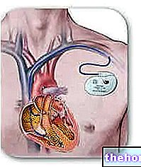
- Defibrillator (ICD). Like the pacemaker, it is also a device implanted under the skin, in this case at the level of the collarbone. It is used when the patient has a tachycardia. normal limit of 100 beats per minute, it emits an electrical discharge directed to the heart.
Since arrhythmic episodes are sometimes due to the onset of particular heart disease, describing the surgical therapy would require a case-by-case analysis. For example, in the face of a valvulopathy such as mitral stenosis, the surgery, aimed at repairing the mitral valve, restores the normal heartbeat. In this case, cardiac arrhythmia is an event resulting from the mitral valve malformation.
On the other hand, it is much simpler to deal with sporadic arrhythmias not linked to other pathologies, therefore not serious: these, in fact, arise after physical exercise, or a strong emotion, and disappear spontaneously without taking antiarrhythmic drugs.If the affected subject takes high quantities of caffeine, the simple correction of the doses taken can solve the problem of cardiac arrhythmia.


