Generality
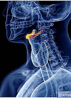
The hyoid bone - or simply hyoid - is an uneven and median bone, shaped like a horseshoe, located in the neck, exactly between the chin and the thyroid cartilage of the larynx.

Numerous muscles are inserted on the upper surface and on the lower surface of the body and horns of the hyoid bone.
The muscles related to the hyoid bone play a fundamental role in the movements of the tongue, pharynx and larynx; therefore, the hyoid bone contributes to the mechanisms of chewing, swallowing, speaking and breathing.
What is the hyoid bone?
The hyoid bone, or simpler hyoid, is the horseshoe-shaped median bone that resides in the neck, at the base of the tongue, exactly between the chin and the thyroid cartilage of the larynx.
The hyoid bone is an element of the human skeleton that is distinguished by its remarkable mobility.

Figure: position of the hyoid bone. As readers can see, when the head is facing forward, the hyoid bone is positioned at the same level as the base of the mandible, anteriorly, and the third cervical vertebra, posteriorly.
ORIGIN OF THE NAME
The term "hyoid" comes from the Greek word "hioeides” (ὑοειδής), whose literal meaning is "ipsilon-shaped". To understand the reason for using the term hyoid, it is appropriate to remind readers that the ipsilon, to which the word refers hioeides, is the small Greek ipsilon, whose shape is very reminiscent of a horseshoe or the vowel "u".
Anatomy
The hyoid bone is an irregular bone, in which it is possible to distinguish a central portion, called the body, and two lateral bony projections, which are called horns.
The hyoid bone has a peculiarity that distinguishes it from all the other bones of the human body: it is the only bone element of the human skeleton that does not articulate with other bones and which is held in position exclusively by a complex of muscles and ligaments.
BODY
The body has the appearance of a sheet arranged transversely, on which it is possible to identify two faces - the front face and the back face - and two margins (or edges) - the upper and lower margins.
- There front face it is convex and represents the site of insertion of the genioioid, mylohyoid, stylohyoid, homohyoid and iogloxy muscles;
- There back face is the concavity resulting from the convex shape of the front face. It establishes relations with the so-called thyroid membrane;
- The top margin it is rounded and inserts part of the thyroid membrane and some fibers of the genioglossus muscles;
- The bottom margin it is, medially, the site of insertion of the sternohyoid muscles and, laterally, the site of insertion of the homohyoid muscles and part of the thyroid muscles.
HORN
The two horns present on each side of the body of the hyoid bone are one longer than the other.
The long horns of the hyoid bone form the so-called pair of major horns, while the short horns of the hyoid bone form the so-called pair of minor horns.
The greater horns they represent the outermost portion of the hyoid bone. With respect to the body, they project in a posterior direction and slightly upwards. Along their course, they tend to become thinner, only to swell again at the extremities, giving rise to a tubercle (the tubercle of the greater horns). The so-called lateral thyroid ligament is inserted on the tubercle of the greater horns.
On each major horn there is one surface - the upper surface - and two margins (or edges) - the medial margin and the lateral margin.
- There upper surface of the greater horns it represents the site of attachment of the ioglossus muscle and part of the digastric and stylohyoid muscles;
- The medial margin of the major horns inserts part of the thyroid membrane and the middle constrictor muscle of the pharynx;
- The lateral margin of the greater horns gives insertion to the thyroid muscle.
Then moving on to minor horns, these are two conical eminences, more internal than the major horns and oriented upwards. They are joined to the body, by means of fibrous tissue, and to the major horns, by means of a "synovial joint.
An important ligament is inserted on the "apex of the minor horns: the stylohyoid ligament."
LIGAMENTS
In summary, the ligaments that have relations with the hyoid bone are: the lateral thyroid ligament and the stylohyoid ligament.
The lateral thyroid ligament is the equal anatomical element, which connects the tubercle of the greater horn of the hyoid bone to the superior horn of the thyroid cartilage.
The stylohyoid ligament, on the other hand, is the even anatomical element, which joins the apex of each minor horn of the hyoid bone to the styloid process of the temporal bone of the skull.
MUSCLES
By convention, anatomists distinguish the muscles that find insertion in the hyoid bone into two broad categories: the muscles that find insertion on the upper surfaces of the components of the hyoid bone (muscles of the upper surface of the hyoid bone) and the muscles that insert on the lower surfaces of the components of the hyoid bone (muscles of the lower surface of the hyoid bone).
The first category includes: the middle constrictor muscle of the pharynx, the ioglossus muscle, the genioglossus muscle, the intrinsic muscles of the tongue and the so-called suprahyoid muscles (digastric, stylohyoid, geniohyoid and mylohyoid).
On the other hand, three of the four subhyoid muscles belong to the second category: the thyroid muscle, the omohyoid muscle and the sternohyoid muscle.
Remember that the muscles that form relationships with the hyoid bone are all equal muscle elements.
VASCULARIZATION
The supply of oxygen-rich blood to the hyoid bone depends on the so-called lingual artery.
Even arterial vessel, the lingual artery originates from the external carotid artery and reaches the hyoid bone, where the greater horn develops.
The lingual artery is important not only because it provides blood supply to the hyoid bone, but also because it gives rise to a branch - the so-called suprahyoid branch - whose job is to supply the muscles of the upper surface of the hyoid bone with blood.
Functions
The hyoid bone is the anchorage site for muscles that allow the movements of the tongue, pharynx and larynx. Therefore, it plays a fundamental role in the physiological functions performed by the aforementioned anatomical structures, namely: in the mechanisms of chewing, swallowing, speaking and breathing.
MOVEMENTS OF THE HYOID BONE DURING SWALLOWING
During swallowing, the hyoid bone moves first upward, then forward, and finally returns to the starting position.
The movement of the hyoid bone depends on the muscles that are inserted into it.
ROLE IN BREATHING
As far as breathing is concerned, the hyoid bone plays a key role in keeping the airways open during nighttime sleep.
Development
The embryonic origin of the hyoid bone sees the second pharyngeal arch and the third pharyngeal arch as protagonists. From the second pharyngeal arch derive the minor horns and the upper portion of the body. From the third pharyngeal arch derive the greater horns and the lower portion of the body.
OSSIFICATION
6 ossification centers contribute to the formation of the hyoid bone: two centers for the body and one center for each horn.
The process of ossification of the hyoid bone begins from the major horns, at the end of fetal development. Shortly thereafter, the ossification of the body begins and, around the 1st-2nd year of life, the ossification of the minor horns begins.
Pathologies
Numerous clinical studies have shown that the hyoid bone is the protagonist of a well known and widespread nocturnal sleep disorder: the so-called obstructive nocturnal apnea syndrome.
Those who conducted the aforementioned studies, in fact, found that, in many people with the aforementioned syndrome, the hyoid bone occupies a lower position than normal and for this reason it conditions the opening mechanism of the airways during sleep.
CAN IT FRACTURE?
The hyoid bone occupies such a position that it is very rare for it to fracture.
This is more true for adults than for children and adolescents, where the hyoid bone is still ossifying (so it is less resistant)
Curiosity
Fracture of the hyoid bone is an event that generally characterizes deaths from strangulation or strangulation


.jpg)


