Cholangiography
If the ultrasound examination is positive, no further investigations are required to confirm the presence of gallbladder stones.If, on the other hand, the ultrasound is negative, further tests may be carried out:
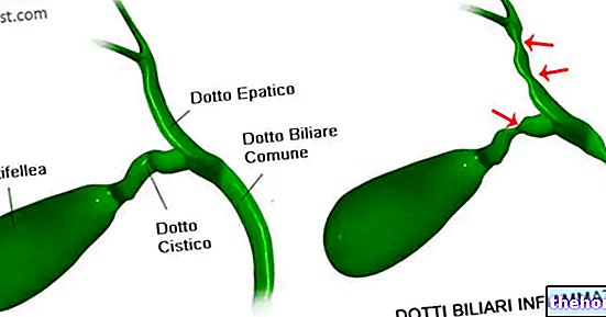
- Endoscopic Retrograde Cholangiography (ERCP): allows to radiologically highlight the state of health of the biliary and pancreatic tracts (choledochus, gallbladder, common hepatic duct, intrahepatic ducts and pancreatic ductal system see: gallbladder anatomy). Contrast medium is injected through a tube inserted orally and made to go down the digestive tract to perform radiograms. Using other catheters it is also possible to perform therapeutic maneuvers such as the extraction of stones or the drainage of bile in case of obstructive jaundice (benign or malignant).
- Percutaneous cholangioscopy (CPT): through a small hole made in the skin of the abdominal wall, a catheter is inserted that directly reaches the biliary tract and injects a radiological contrast medium. Obviously, because of this incision, percutaneous cholangioscopy is a fairly invasive examination which is used only if the previous technique (ERCP) is contraindicated. Precisely for this reason, percutaneous cholangioscopy must be performed in specialized centers that can intervene by removing the stones present in the biliary tract.
- NMR-cholangiography: is an innovative technique that exploits the potential of nuclear magnetic resonance (NMR). It allows the computerized reconstruction of the intrahepatic biliary tract, allows the visualization of stenosis and lithiasis and is free of side effects. The only flaw lies in the difficult interpretation of the images and the impossibility of removing any obstacles to the outflow of bile (stones).
A normal direct radiograph of the abdomen is able to visualize only the radiopaque stones (it can visualize the pigment stones quite well but not those rich in cholesterol).
Care and Treatment
Read also: Remedies for Gallstones
If "liver" stones are discovered occasionally and are not causing symptoms, the best thing to do is not to worry. The probability of developing biliary colic in the following year is in fact very low (in the order of 2-3%). The risk of tumor formation inside a gallbladder affected by stones exists but it is overall very low, so there is no need to worry too much even about this eventuality.
If the gallbladder stones have already caused biliary colic, the chances of this colic recurring are rather high (about 60% over the next two years). For this reason, after colic or other complications, the indication main is to intervene surgically by removing the gallbladder (cholecystectomy).
Cholecystectomy
In recent years, the use of this intervention is increasingly made of a preventive nature, especially if the calculations are small and multiple. The risk that these pebbles move causing the typical complications of lithiasis (presence of stones in the gallbladder), even if quite low, exists; consequently, a prophylactic approach to the disease is certainly preferable to emergency surgery.

Pharmacological alternatives
Cholecystectomy surgery is the only possibility to definitively solve the problem. In fact, there are several medical therapies capable of destroying cholesterol stones through drugs similar to bile salts, but generally take a very long time and above all do not prevent the reappearance of gallbladder stones.
For further information: Medicines for the treatment of gallbladder stones
How it is done
Thanks to the introduction of videolaparosurgery, known to most as a "minimally invasive" technique, the treatment of gallbladder stones has undergone considerable modernization in recent years. Special instruments are inserted through small incisions made in the patient's abdomen. they will be maneuvered by the surgeon with the aid of the images coming from a micro-camera introduced at the umbilical level. The introduction of gas into the abdominal cavity helps to lift the wall of the abdomen making operations easier.
Thanks to this type of surgery, the post-operative course is faster and the patient can be discharged already 1-3 days after the operation, without the pains and difficulties of recovery typical of the past.
In general, after the removal of the gallbladder, life resumes normally. In the postoperative phase a tendency to diarrhea may occur, but the organism quickly adapts and these problems disappear.
To learn more, read the article on Cholecystectomy
Other articles on "Liver Stones - Diagnosis and Treatment"
- Biliary colic and complications
- Gallbladder stones, gallbladder stones
- Risk factors, symptoms and complications
- Gallbladder Stones - Medicines to Treat Gallbladder Stones
- Nutrition and Gallstones
- Diet and Gallbladder Stones

