So is the Cornea
The cornea is the membrane that covers the front of the eye, through which it is possible to see the iris and the pupil.
Transparent and avascular, this structure represents the first "lens" that light encounters on its way to the brain.The cornea is, in fact, an essential element of the ocular dioptric system: it allows the passage of light rays towards the internal structures of the eye and helps to focus the images on the retina.
The cornea is made up of superimposed layers, the outermost of which is the stratified paving epithelium, while the subsequent ones are formed by a dense interweaving of collagen fibrils arranged in lamellae, with a glycoprotein matrix that unites them and makes them transparent.

Appearance and structure
The cornea forms the anterior portion of the fibrous layer of the eyeball. The sclera - that is the "white part of the" eye "with which the corneal surface is structurally in continuity - represents, on the other hand, the five posterior sixths of the same tunic.
The external surface of the cornea is convex and has a slightly elliptical shape, with the horizontal diameter greater than the vertical one. The inner face, on the other hand, is concave and has approximately the same radius of curvature as the anterior part (the anterior radius of curvature is equal to 7.2 mm, while the posterior one is 6.8 mm). thin in the central area (about 520-540µm) compared to the periphery (about 0.7-0.8 mm).
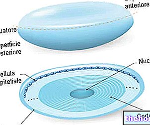
From a structural point of view, in the cornea there are five layers (from the outside towards the inside):
- Corneal epithelium: multi-layered paving type, it is 50-60 µm thick (about one tenth of the total thickness of the cornea). Arranged in 5-6 layers there are basically three types of cells: basal, polygonal (intermediate) and superficial flat, which represent different maturation stages of the same cell unit. These elements, with an optically perfect shape, are joined together by tight joints. The basal cells are endowed with high replicative activity, protect the ocular surface from mechanical abrasion and form a permeable barrier.
- Bowman's lamina (or anterior limiting membrane): placed under the corneal epithelium, it is a cell-free membrane consisting of an interweaving of collagen fibers, immersed in a matrix of proteoglycans (thickness: 10-12 µm).
- Corneal stroma: constitutes most of the total thickness of the cornea (400-500 µm); it is mainly made up of connective fibers, glycoprotein matrix and keratocytes. In the stroma, type I collagen fibrils organize themselves into different lamellar layers, spacing out from each other with extreme precision. Keratocytes combine to form a sort of network between one lamellar layer and the next. The precise three-dimensional arrangement of the corneal fibers and cells, together with the identical refractive index of the matrix interposed between the stromal lamellae, are responsible for the perfect transparency of the cornea.
- Descemet's membrane (or posterior limiting membrane): like Bowman's lamina, this layer is acellular and formed by a thin network of collagen fibers, radially arranged; it has a variable thickness of 4-12 µm (it tends to thicken proportionally to age).
- Endothelium: it is the deepest layer of the cornea, consisting of a single layer of flattened hexagonal-shaped cells, rich in mitochondria, connected by desmosomes and intercellular densities. The endothelium plays an important role in regulating the exchanges between the aqueous humor and the upper layers of the cornea; moreover, it maintains the tropism and corneal transparency.
Dua layer
In 2013, during a scientific research aimed at clarifying some aspects about the outcome of corneal transplants, a sixth corneal layer, called the "Dua layer", was identified.
Located in the posterior part of the cornea, between the stroma and Descemet's membrane, the Dua layer is only 15µm thick. This can only be highlighted through the examination with the electron microscope, after the insufflation of tiny air bubbles, which gently induce the separation of the different layers that make up the cornea.
Despite the very thin thickness, the Dua layer is extraordinarily resistant (it can withstand pressure values of 1.5-2 bar). According to the authors of the study, if the surgeons were able to inject a bubble near the layer of Dua, the risk of injury secondary to corneal transplantation could be reduced, thanks to the high degree of resistance of this membrane. Furthermore, the results of this research can help to understand numerous corneal diseases, including acute hydrops, descemetocele and pre-Descemet dystrophies.

-corpi-estranei-e-altre-cause.jpg)
.jpg)
