Generality
The exophthalmos consists in the protrusion of the eyeball, anteriorly outside the orbit; as a result of this anomaly the eyes become visually "protruding" or prominent.
The terms exophthalmos and proptosis are often used interchangeably, but with some reservations:
- Exophthalmos is used to indicate bulging eyes in endocrine-related conditions;
- Proptosis indicates, more precisely, the protrusion of the eyeballs caused by other causes (orbital tumor, vascular pathologies, retrobulbar haemorrhage, etc.)
.jpg)
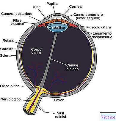
The protrusion of the eye is secondary to the increase in the orbital volume within the bone boundaries, which instead remain fixed. The orbit is in fact closed at the posterior, medial and lateral walls; therefore, any enlargement of the structures located inside it will cause the anterior displacement of the eyeball, with consequent exophthalmos.
Causes
The exophthalmos can represent the result of many processes deriving from primary orbital pathology (isolated or proximal) or from systemic diseases. The etiological basis can be mainly inflammatory, vascular, neoplastic or infectious. In adults, thyroid orbitopathy (ie the pathology eye socket of thyroid origin) is the most common cause of unilateral and bilateral exophthalmos. In particular, Graves' disease - an autoimmune disease that causes hyperthyroidism - is often associated with exophthalmos: bulging eyes are due to abnormal infiltration of lymphocytes, plasma cells and mast cells at the level of the orbital connective tissue; this determines a deposition of collagen and glycosaminoglycans in the extrinsic muscles of the eye, which in turn leads to fibrosis and a further enlargement of the orbital volume.
Proptosis is sometimes associated with the onset of tumors that develop in the eye socket. The complete or partial dislocation of the orbit is also possible due to direct trauma or swelling of the surrounding tissue. In children, unilateral exophthalmos is commonly caused by orbital cellulitis, while neuroblastoma and leukemia are likely if the condition is bilateral.
The main causes of exophthalmos and proptosis are shown in the table.
Neoplastic
Graves' ophthalmopathy
Orbital cellulite
Dacryocystitis
Mucormycosis
Orbital inflammatory syndrome
Wegener's granulomatosis
Leukemia
Meningioma
Nasopharyngeal angiofibroma
Hemangioma
Lacrimal gland adenoma
Glioma
Vascular
Other causes
Carotid-cavernous fistula
Aortic insufficiency
Cavernous sinus thrombosis
Hyperthyroidism
Neuroblastoma
Dermoid cyst
Frontal Sinus Mucocele
Orbital and facial fracture
Retrobulbar hemorrhage
Progeria
Symptoms
The most obvious clinical sign is the anterior displacement of the eye from the orbit.
Exophthalmos can be accompanied by other symptoms:
- Eyelid swelling: can be associated with conjunctival chemosis (protrusion of the bulbar conjunctiva with respect to the underlying tissues) and blepharoptosis.
- Difficulty closing the eyelids completely while blinking or sleeping at night;
- Double vision: caused by the restriction of the movement of the extrinsic muscles of the eyeball, which can be the source of inflammation (myositis) or be compressed by a growing tumor.
- Redness and pain: tend to occur in the presence of inflammation, infection or a rapidly progressing tumor. In severe cases, there may be secondary exposure keratopathy, as a result of incomplete closure of the eyelid on the cornea. Corneal surface compromise can cause pain and affect visual acuity.
- Decreased vision: The patient may experience decreased vision. Visual acuity may be impaired by direct involvement of the optic nerve in the pathophysiology of exophthalmos or if the macula is distorted by a lesion that pushes behind the globe (tumor or hemorrhage).
Depending on the cause, other eye symptoms may be present. If exophthalmos is caused by a thyroid-related condition, such as Graves' disease, in addition to bulging eyes, the following can also occur:
- Eye inflammation, redness and pain;
- Dry eye
- Excessive tearing
- Sensitivity to light (photophobia).
Complications
Particularly severe proptosis can cause lagophthalmos (failure to close the eyelids). Continuous exposure of the eye can cause dryness and possible corneal damage (infections or ulcers), due to the increased friction during blinking. The pathological process that causes the displacement of the eyeball can also compress the optic nerve or ophthalmic artery, causing blindness. Other possible complications include conjunctivitis and optic atrophy. Exophthalmos can increase the pressure behind and inside the eye (intraocular pressure). Excessive intraocular pressure increases the risk for other eye diseases, such as glaucoma. If a person suddenly develops proptosis, especially in one eye, a very serious problem may be present, which should always be evaluated immediately by an ophthalmologist.
Diagnosis
The exophthalmos is often easy to recognize by the evident protrusion of the eyeballs.
A thorough medical history of the patient is the key to establishing a diagnosis. The clinical presentation, in fact, varies according to the underlying cause. However, the very nature of exophthalmos results in some common features. The direction of the protrusion, severity, rate of onset, and associated symptoms often give a good indication as to the underlying cause, but this usually has to be be confirmed with further investigation. The ophthalmologist will check the range of eye movements, visual acuity, pupillary function, visual field defects and the width of the interpalpebral fissure. Measurement of the degree of exophthalmos is done using an instrument called an exophthalmometer. Most sources define proptosis as an eyeball protrusion greater than 18 mm. Blepharoptosis and lagophthalmos (incomplete closure of the eyelids) are additional signs to be considered during the examination.
Palpation of the anterior orbit allows for assessment of the level of swelling, consistency and mass mobility. Edema may denote an inflammatory process or "neural invasion by neoplasia." Tactile inspection of the globe may reveal pulsations secondary to communications arteriovenous. If cancer is suspected as a cause of proptosis, computed tomography (CT) or magnetic resonance imaging (MRI) may be done to examine the eye socket in more detail. The results should point to further laboratory studies. For example, in the case of lymphoma, hematology studies, body imaging and a bone marrow biopsy may be indicated. In patients with orbital cellulitis, cultures of blood and nasal samples and a complete blood count can be done. Blood tests or a thyroid function test allow you to check if the thyroid gland is functioning properly.
Treatment
Treatment depends on the underlying cause. Once the etiology of exophthalmos or proptosis has been established, medical therapies will be directed at reversing the underlying problem and minimizing ocular complications. Meanwhile, artificial tears can be used to provide symptomatic relief and protect the exposed cornea. For more severe cases, surgery may be required. Patients should be monitored regularly to assess the degree of exophthalmos and the complications resulting from this eye disease. Additionally, any corneal damage should be identified early and resolved.


