What is a hemangioma?
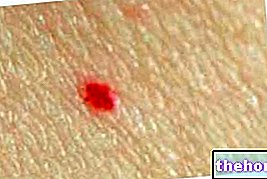
A hemangioma is a benign tumor of the endothelial cells, which normally line blood vessels. The lesion is characterized by a dense collection of capillaries, which forms a superficial or deep lump. Hemangiomas can occur anywhere on the body, including the layers of the skin and internal organs.

Causes
The cause is currently unknown, however several studies have suggested the intervention of estrogens in the proliferation of the hemangioma. In particular, hypoxia (lack of oxygen) localized to soft tissues, associated with the increase in the levels of circulating hormones after birth, may be a stimulus for the onset of benign tumor. Some scientific evidence suggests, however, a role of placental tissue during gestation Other theories have been proposed, but more research is needed to confirm the causes of the development of hemangiomas.
In hemangiomas, endothelial cells multiply at a very rapid rate. Under the microscope, the lesions consist of clusters of thin blood vessels with endothelial lining and separated by scant connective tissue. Glut1 is a highly specific histochemical marker for hemangioma and can be used to differentiate it from vascular malformations.
Emagioma vs. vascular malformation
The terminology used to describe and classify vascular abnormalities and nodules consisting of blood vessels has changed over time. The term "hemangioma" was originally used to describe any tumor-like vascular lesion, congenital or late onset. In fact, it is possible to distinguish these conditions into two families: 1) self-involutional tumors and 2) malformations present at birth and substantially stable. This distinction makes possible an early differentiation, between lesions that tend to resolve spontaneously (hemangioma) and permanent ones (vascular malformations).
Emagioma
Vascular malformation
Histology
High proliferation of endothelial cells.
Normal cell phone turn-over.
Presence at birth
Usually absent.
Present (not always evident).
Clinic
Noticeable a few weeks after birth. The proliferative phase continues for 1-2 years, then the hemangioma spontaneously involves.
It grows proportionally with the child's development.
Diagnosis
History and clinical signs.
Imaging (MRI, CT and angiography).
Treatment
Observation; if the hemangioma is large, in anatomically sensitive areas and if involution is not complete, it can be treated with corticosteroids or surgically.
It depends on the location, size, symptoms, etc. Treatment options include sclerotherapy with or without excision and surgery.


.jpg)


