Generality
The pelvis, or pelvis, is the lower part of the human body, between the abdomen, above, and the thighs, below.
The pelvis includes: the bones of the pelvis, which form a structure also known as the pelvic girdle; the pelvic cavity, which is the space enclosed by the pelvic girdle; the pelvic floor, which is basically the base of the pelvic cavity; finally, the perineum, which is the anatomical area under the pelvic floor.

The female pelvis has some differences from the male pelvis, especially as regards the arrangement of the pelvic bones and the internal space that these bones create (pelvic cavity). These differences are related to reproduction and to the fact that the female pelvis is the site of development and growth of the fetus.
The pelvis has three important functions: it supports and, at the same time, loads the weight of the upper body on the lower limbs; it hosts joints and muscles fundamental to locomotion and upright posture; finally, it encloses and protects organs such as the bladder, the urethra, the rectum, the uterus (in women), the ovaries (in women), the fallopian tubes (in women), the prostate (in men) ) etc.
What is the pelvis?
The pelvis, also known as the pelvis or pelvic region, is the lower part of the human body, placed precisely between the abdomen (in the upper position) and the thighs (in the lower position).
Anatomy
The pelvis includes:
- The bones of the pelvis (or pelvic bones);
- The pelvic cavity, space resulting from the particular arrangement of the pelvic bones;
- The pelvic floor, which forms the basis of the pelvic cavity;
- The perineum, located below the pelvic cavity.
BONES OF THE PELVIS
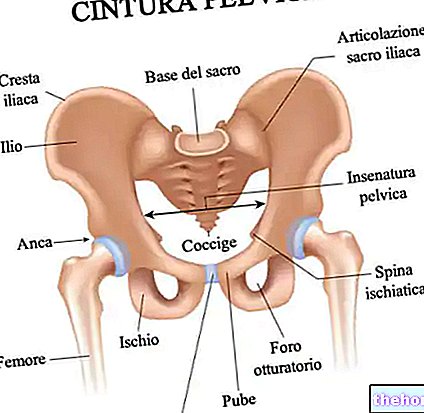
The bones of the pelvis are 4: the sacrum, the two iliac bones and the coccyx.
In relating to each other, the bones of the pelvis give life to an anatomical structure with an oval shape, which experts define as the pelvic girdle. The pelvic girdle represents the connection between the so-called axial skeleton (consisting mainly of the skull, rib cage and spine) and the lower limb skeleton.
Analyzing briefly the various bones of the pelvis, the sacrum and the coccyx represent the posterior portion of the pelvic girdle as well as the two terminal segments of the vertebral column, in which an organ essential to life such as the spinal cord is housed. The iliac bones, on the other hand, represent the lateral portions and the anterior portion of the pelvic girdle; they constitute in effect the hips, include the hip joints and, joining on the anterior portion of the pelvic girdle, form the so-called pubic symphysis. These can be divided into three regions, known as the ilium , ischium and pubis, the iliac bones, are connected to the sacrum and from here they develop, according to the modalities just mentioned.
From a functional point of view, the bones of the pelvis have two main tasks: supporting the weight of the upper body and connecting the latter to the lower limbs (specifically to the two femurs, through each hip).
The support function, towards the upper body, is especially important when the human being stands up, sits, walks, runs, etc.
The binding function with the lower limbs, on the other hand, is essential for walking.

- The two sacro iliac joints: these are the joint elements that connect the sacrum to the two iliac bones.
- The lumbo-sacral joint: it is the joint element that connects the last lumbar vertebra with the first sacral vertebra.
- The sacro-coccygeal joint: it is the joint element that connects the last sacral vertebra with the first coccygeal vertebra.
Joints of each hip bone:
- The sacrum iliac joint.
- The pubic symphysis: it is the joint that unites each iliac bone in the front.
- The hip joint: it is the joint element that connects the iliac bone to the femur.
Joints of the coccyx:
- The sacro-coccygeal joint.
PELVIC CAVITY
The pelvic cavity is the body cavity, bounded by the pelvic girdle, anteriorly, posteriorly and laterally, by the pelvic floor, below, and by the so-called pelvic entrance, above.
Between the abdomen and the perineum, the pelvic cavity has a characteristic funnel shape.
Inside the pelvic cavity, large arteries, veins, muscles, nerves and very important organs (the so-called pelvic organs) take place, including:
- The bladder, located just behind the pubic symphysis;
- The rectus intestine, located approximately in the center of the posterior part of the pelvis, immediately in front of the borderline between the sacrum and the coccyx;
- The sigmoid colon (or sigmoid colon), located to the left of the rectum and communicating with the latter.
- The uterus, fallopian tubes, ovaries and vagina in women;
- The prostate, the vas deferens and the seminal vesicles in man.
Most anatomical descriptions of the pelvic cavity report that the pelvic cavity can be divided into two regions: an upper one, called the large pelvis or false pelvis, and a lower one, called the small pelvis or true pelvis.
The large pelvis is a space shared with the abdominal cavity; the small pelvis, on the other hand, is the actual pelvic cavity, the one that includes the aforementioned organs.
PELVIC FLOOR
Also known as the pelvic diaphragm, the pelvic floor is a "rhomboid" area, consisting predominantly of muscle tissue and to a lesser extent connective tissue, which extends from the pubic symphysis to the coccyx.
The pelvic floor - especially its muscle tissue - has two important functions: one function is to close the pelvic cavity below and to support the load of the pelvic and abdominal organs; the other function, on the other hand, is to control the openings towards the outside of the rectum and urogenital organs, by means of special perforations on the muscle tissue and connective tissue (the so-called rectal hiatus and urogenital hiatus).
In essence, therefore, the pelvic floor must guarantee a "supportive action, against the bladder, rectum, organs of the reproductive system, etc., and, at the same time, must ensure the passage, through its constituent tissues, of those anatomical structures that open outwards.
The muscle tissues of the pelvic floor belong to two very important muscles, certainly known to most, which are: the levator anus muscle and the coccygeal muscle.
PERINEUM
In the human being, the perineum is the lozenge-shaped anatomical area corresponding to the lower extremity of the pelvis.
Underlying the pelvic floor, it traces the extension from the pubic symphysis to the coccyx.
Looking at it from the outside, it is the area of the human body which, in the coccygeal-pubic direction, goes from the anus to the genital organs (vulva, in women, and testicles, in men) and which, in a transverse direction, is between the two thighs.
The perineum includes several muscles (including the external anal sphincter muscle, the urethral sphincter muscle, the bulbospongiosus muscle, the superficial transverse perineal muscle, and the deep transverse perineal muscle), connective tissue, collagen fibers, skin tissue, subcutaneous tissue, and ligaments.
According to the most traditional anatomical descriptions, it can be divided into two semi-triangles having the base in common: the so-called urogenital triangle, anteriorly, and the so-called anal triangle, posteriorly.
The urogenital triangle contains the genital organs, while the anal triangle encloses the anus.
The junction point of the two triangles - that is, where the base resides in common - coincides with the position occupied by a fibromuscular structure, called the perineal body. The perineal body is a very important anatomical element for at least two reasons:
- It has the task of maintaining the integrity of the pelvic floor, avoiding the prolapse of organs such as the rectum, the bladder, the uterus (in women) and so on.
- It is used to hook the levator ani muscle (belonging to the aforementioned pelvic floor) and the muscles attributable to the perineum, ie the external anal sphincter muscle, the urethral sphincter muscle, the bulbospongiosus muscle and the two transverse perineal muscles, superficial and deep.
Thanks to the innervation of the so-called pudendal nerve, the perineum represents an erogenous zone, both for the male and for the female gender.
DIFFERENCES BETWEEN MAN AND WOMAN
The female pelvis has several differences from the male pelvis. These differences are essentially linked to reproduction and to the fact that the pelvic cavity of women is responsible for hosting the fetus and facilitating its escape at the time of delivery.
In the "list of differences existing between the female pelvis and the male pelvis, the following certainly cannot be missing:
- The presence, in the woman, of the uterus, fallopian tubes, ovaries and vagina, fundamental organs for the development of the fetus;
- The greater size and width of the female pelvis, compared to the male one, which is not only narrower, but also taller and more compact;
- The greater diameter of the female pelvic inlet, compared to the male one;
- The greater thickness and greater heaviness of the male iliac bones;
- The greater length and narrowness of the male sacral bone as well as the presence, in the latter, of a wider sacral promontory (N.B: the sacral promontory is the bony prominence that articulates the first sacral vertebra with the last lumbar vertebra);
- The greater acuity, in man, of the angle created by the two lower branches of the pubic bones. In the male, the aforesaid angle is about 70 °; in the female, on the other hand, it is between 90 and 100 °;
- The greater distance, in women, between the acetabula (N.B: the acetabulum is the concavity of the iliac bone which houses the head of the femur and forms the hip joint).
If the female pelvis were like the male pelvis (therefore narrower than it actually is), the fetus could not grow adequately and women, as the pregnancy progressed, would have more and more difficulty walking.
Fortunately, evolution has come up with a solution to these drawbacks.
Functions
The pelvis fulfills at least 3 important functions:
- It supports the weight of the upper body and, at the same time, discharges it from the axial skeleton to the lower limb skeleton (which represents a part of the so-called appendicular skeleton);
- It forms joints (eg: hip) and inserts muscles (eg: some muscles of the hip), both of which are essential for locomotion and the maintenance of upright posture;
- It encloses and protects, both through bone structures (the pelvic girdle) and through a solid network of muscles (the so-called abdominals), the so-called pelvic organs (bladder, urethra, rectum, sigma, reproductive organs, etc.).
Muscles of the hip that have relations with the bones of the pelvis:
- Gluteal muscle group
- Gluteus maximus muscle
- Gluteus medius muscle
- Gluteus minimus muscle
- Tensor fascia lata muscle
- Adductor muscle group
- Adductor brevis muscle
- Adductor longus muscle
- Adductor major muscle
- Pectineus muscle
- Gracilis muscle
- Iliacus muscle
- Lateral rotator muscle group
- Internal obturator muscle
- External obturator muscle
- Piriformis muscle
- Superior twin muscle
- Lower twin muscle
- Square muscle of the femur
- Others
- Rectus femoris muscle
- Sartorius muscle
Clinic
From a clinical point of view, the pelvis is very interesting, for at least two reasons: because its bone component is often subject to fractures and because it is the protagonist of a particular painful sensation which, in medical jargon, is called pelvic pain.
BONE FRACTURES
Fractures of the pelvic bones, known more simply as fractures of the pelvis, are injuries of generally traumatic origin, which can also affect more than one bone element.
The typical symptom of pelvic fractures is pain where the fractured bone resides; less frequent but still important symptoms are: lameness (the degree varies according to the severity and location of the fracture), swelling and presence of hematoma.
The treatment of pelvic fractures depends on the severity of the bone lesion present: for less severe fractures, conservative therapy (or non-surgical therapy) is sufficient, which includes rest until the bone has been welded, the use of aids for the walking and the intake of painkillers and anticoagulants; for major fractures, however, surgical therapy is essential, followed by an appropriate period of rest.
CAUSES OF PELVIC PAIN
The possible causes of pelvic pain are numerous, especially among women. For this reason, to simplify consultation, doctors and experts have deemed it appropriate to divide them into at least two broad categories, which correspond to:
- The category of gynecological causes, exclusive to the female sex, e
- The category of non-gynecological causes, of which both men and women can be victims;
Among the gynecological causes of pelvic pain, certainly deserve a mention: dysmenorrhea (or painful menstruation), ovulation, endometriosis, rupture of an ovarian cyst, degeneration of a uterine fibroid, episodes of ovarian or tubal torsion. , vulvodynia, uterus prolapse, pelvic inflammatory disease, spontaneous abortion, episodes of ectopic pregnancy and rupture of a tubo-ovarian abscess.
Among non-gynecological causes of pelvic pain, on the other hand, there are: episodes of gastroenteritis, inflammatory bowel disease, appendicitis, diverticulitis, tumors of the rectum or sigmoid intestine, constipation, intestinal obstruction , perirectal abscess, irritable bowel syndrome, urinary tract diseases (such as cystitis, pyelonephritis, urolithiasis and tumors), episodes of intestinal perforation and stretching of the abdominal muscles that contain the organs of the pelvis.

