Generality
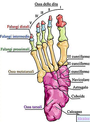
The bones of the foot are, in the human being, the skeletal structure of the terminal tract of each lower limb. They are a total of 26 and, according to anatomists, can be divided into three large groups: the tarsal bones (or tarsal or tarsal bones) , the metatarsal bones (or metatarsals) and the bones of the toes (or phalanges of the foot).
The bones of the foot have a supporting function, they allow the human being to be a bipedal animal, they form a series of very important joints for the functionality of the foot and, finally, they insert tendons which are fundamental for locomotion.
Like any bone in the human skeleton, the bones of the foot can also fracture.
What are the bones of the foot?
The bones of the foot are, in the human being, what constitutes the skeleton of the terminal part of each lower limb.
Inside the human body, the feet are two fundamental anatomical structures:
- Ensure stability in the standing position;
- Absorb a large part of the weight of the body;
- Allowing locomotion. Without the feet, the human being would not be able to walk, run, jump and so on.
Anatomy
In all 26, the bones of the foot can be divided into three large groups: the bones of the tarsus (or more simply the tarsus), the metatarsal bones (or metatarsals) and the bones of the toes (or phalanges of the foot).
The bones of the tarsus are 7 and represent the proximal portion of the skeleton of the foot; the metatarsal bones are 5 and represent the intermediate portion of the foot skeleton; finally, the phalanges of the foot are 14 and represent the distal portion of the foot skeleton.
In anatomy, proximal and distal are two terms with opposite meanings.
Proximal means “closer to the center of the body” or “closer to the point of origin.” Referring to the femur, for example, it indicates the portion of this bone closest to the trunk.
Distal, on the other hand, means "farther from the center of the body" or "farther from the point of" origin. "Referred (again to the femur), for example, it indicates the portion of this bone farthest from the trunk (and closest to the "knee joint).
BONES OF THE TARSO
The tarsal bones, also known as the tarsal bones, are irregularly shaped bones that form a compact structure located between the distal ends of the tibia and fibula and the proximal ends of the metatarsal bones.
The foot bones that form the tarsus are: the talus, the calcaneus, the navicular bone, the cuboid bone, the lateral cuneiform bone, the intermediate cuneiform bone and the medial cuneiform bone.
- The talus and the calcaneus represent the most proximal bones of the tarsus and play a fundamental role in the formation of the ankle, ie the joint that allows dorsiflexion, plantarflexion, eversion and inversion of the foot.
In this case, the talus takes place, with its upper margin, inside the concavity deriving from the particular anatomy of the distal extremities of the tibia and fibula; this concavity is called a mortar.
The calcaneus, on the other hand, participates in the ankle joint by inserting some extremely important ligaments for the correct functioning of the aforementioned joint element; the ligaments in question are the tibio-calcaneal ligament and the calcaneofibular ligament.
Together, the talus and calcaneus make up the back of the foot (or hindfoot). - The navicular is the intermediate bone of the tarsus; it resides anterior to the talus, posterior to the three cuneiforms and lateral to the cuboid. It has a protuberance, which serves to insert a tendon, called the posterior tibial tendon.
- The cuboid and the three cuneiforms are the most distal bones of the tarsus.
With its cube-like appearance, the cuboid bone occupies a lateral position with respect to the three cuneiforms and borders the calcaneus, posteriorly, and the two outermost metatarsal bones (fourth and fifth metatarsal), anteriorly.
With a wedge-like appearance, the three cuneiforms (lateral, intermediate and medial) reside in front of the navicular bone and behind the three innermost metatarsals (first, second and third metatarsal).
The particular arrangement of the three cuneiforms and the cuboid allows the neighboring metatarsal bones to form the so-called transverse arch of the foot.
METATARSAL BONES
The metatarsal bones, or metatarsals, are long bones, arranged parallel to each other, in which it is possible to distinguish three regions: a central region, called the body; a proximal region, called the base; finally, a distal region, identified with the term head.
The base of the metatarsals borders on the bones of the tarsus: starting from the inner side of the foot, the first three metatarsals adhere, one each, to one and only one of the three cuneiforms (the first metatarsal to the medial cuneiform, the second metatarsal to the intermediate cuneiform and the third metatarsal to the lateral cuneiform), while the last two metatarsals (fourth and fifth metatarsal) adhere to the cuboid bone.
The head of each metatarsal borders the first phalanx of each toe: as a result, each metatarsal corresponds to a toe.
Between the base of the metatarsals and the bones of the tarsus are a series of joints, as well as between the head of the metatarsals and the first phalanges of the foot.
FOOT MUDS
Cylindrical in shape, the phalanges of the foot are the skeleton of the 5 toes.
Except the first toe - the only one made up of 2 phalanges - all the other toes have 3 phalanges each.
The phalanges closest to the head of the metatarsals are called first phalanges (or proximal phalanges); starting from these, the following ones are called second phalanges (or intermediate phalanges) and third phalanges (or distal phalanges).
Between each phalanx there is a "joint", which gives the fingers a certain mobility.
Note: in the first toe, the numbering of the phalanges ends with the second phalanges.
Function
The bones of the foot have a supporting function, allowing the standing position on two limbs; they form joints that are fundamental to the functionality of the foot; they give insertion to ligaments which are an integral part of the aforementioned joints; finally, they insert very important tendons for locomotion, such as the Achilles tendon.
Pathologies
Like all bones in the body, the bones of the foot can also fracture.
There are three types of fractures affecting the bones of the foot: fractures of the tarsal bones, fractures of the metatarsals (or metatarsal fractures) and fractures of the phalanges.
FRACTURE OF A TARSTUS BONE
Fractures of the foot bones located in the tarsus can be traumatic in nature (most cases) or due to excessive stress (minority of cases).
Among the bones of the tarsus most prone to traumatic fractures are the talus and the calcaneus.
The navicular bone and once again the calcaneus are among the tarsal bones most prone to stress fractures.
Individuals who are victims of a traumatic tarsal fracture must wear a cast - clearly on the fractured foot - for at least 6 weeks and avoid, during this time, putting weight on the limb with the fracture.
Those who are victims of a stress fracture to the tarsal bones can limit themselves to the use of a brace or a crutch, to limit the weight borne by the tarsus during walking.
Typical clinical manifestations of a tarsal bone fracture are foot pain and lameness.
For an accurate diagnosis, X-ray examination of the sore foot, physical examination and medical history are essential.
FRACTURE OF THE METATARSUS
The metatarsals are bones in the foot that can be fractured in at least three different ways:
- As a result of a violent blow, directed on the back of the foot. This is the case, for example, of a heavy object that falls on the foot.
Fractures of the metatarsals due to violent impacts are the most common. - As a result of a stress factor that affects the foot in general or a part of it in particular. This type of fracture is called a metatarsal stress fracture and mainly affects the metatarsals of the 2nd, 3rd and 4th toes. It is very common among high-level athletes and is usually a microfracture.
- As a result of an excessive inversion of the foot. With a violent and very marked inversion of the foot, the peroneus brevis muscle could “pull” the metatarsus of the 5th finger and cause it to break.
Typical clinical manifestations of a metatarsal fracture are: fractured foot pain and lameness.
For a certain diagnosis, an X-ray examination of the foot is essential.
The treatment of metatarsal fractures varies according to the location of the bone break and whether the latter is compound or displaced.
In fact, in certain cases, rest and immobilization of the lower limb may be enough; in others, however, surgery aimed at welding the bone fracture may be indispensable.
FRACTURE OF A PHALANGE OF THE FOOT
Fractures of one or more phalanges of the foot are conditions of mild severity, which arise as a result of traumatic events to damage to the toes. In general, the treatment of fractures affecting the bones of the foot making up the toes consists of a rest period of 20-30 days.






.jpg)


