Generality
The ear is the organ that allows the perception of sounds (the so-called sense of hearing) and that guarantees the static and dynamic balance of the body.
Divided into three compartments - whose names are external ear, middle ear and inner ear - the ear is made up of portions of a cartilage nature, bones, muscles, nerves, blood vessels, sebaceous glands and ceruminous glands.

In the external ear, the main elements are: the auricle, the external auditory canal and the lateral surface of the eardrum; in the middle ear, the most important elements are: the eardrum, the three ossicles, the Eustachian tube, the window oval and round window; finally, in the inner ear, the most relevant elements are: the cochlea and the vestibular apparatus.
What is the ear?
The ear is the organ of hearing and balance.
In humans and mammals in general, the ear has three components, which anatomists call: outer ear, middle ear and inner ear.
Anatomy
The ear is an even organ, which resides at the level of the head.
It includes portions of a cartilage nature, bones, muscles, nerves, arterial vessels, venous vessels, sebaceous glands and ceruminous glands.

External ear
The outer ear is essentially the component of the ear visible to the naked eye on the sides of the head. The main parts that make it up are: the auricle, the external auditory canal (or external acoustic meatus) and the external face of the eardrum (or tympanic membrane).
- Auricle. Covered with skin, it is a predominantly cartilage structure, on which anatomists identify various characteristic areas, including: two curved rhymes, one more external than the other, called helix and antihelix; two protrusions, called tragus and antitragus, which tend to cover the external acoustic meatus, the concha, which is the concave region in which the opening of the external auditory canal takes place; finally, the lobe, made up of adipose tissue and located on the lower edge.

- External auditory canal. Between 2.5 and 4 centimeters long and covered with skin, it is the canal that, with a characteristic S-curve, goes from the auricle (precisely from the hollow) to the eardrum.
The initial tract of the external auditory canal is cartilaginous in nature, while its final tract is bony in nature. The bony portion that constitutes the final tract belongs to the temporal bone of the skull and is called the auditory bubble (or tympanic bubble).
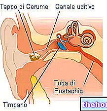
The skin that lines the external ear canal is rich in sebaceous glands and ceruminous glands. The glands' job is to secrete substances such as earwax, which serve to protect the ear in general from potential threats. - External face of the eardrum. It is the face that looks in the direction of the opening of the external ear canal.
On the outer ear there are various muscles and ligaments.
Distinguished into extrinsic and intrinsic, the muscles of the human external ear are structures that are almost completely irrelevant from the functional point of view.
On the contrary, the ligaments have a role of some importance: those defined extrinsic connect the cartilage to the temporal bone, while those defined intrinsic keep the cartilage in place and give shape to the auricle.
Middle ear
The middle ear is the component of the ear between the outer ear and the inner ear. Its main constituent parts are: the tympanic membrane (or eardrum), the tympanic cavity, in which the so-called three ossicles take place, the auditory tube, the oval window and the rounded window.
- Tympanum. Located at the end of the external auditory canal and immediately before the tympanic cavity, it is a thin transparent oval-shaped membrane, which has the task of transmitting the sound vibrations, penetrated through the external ear, to the chain of the three ossicles.
The tympanic membrane can be divided into two regions: the so-called pars flaccida and the so-called pars tensa.
Very often anatomists describe it as the border point between the outer ear and the inner ear. - Tympanic cavity. Also known as the eardrum cavity or tympanic cavity, it is a hollow area that originates at the level of the so-called petrous rock of the temporal bone of the skull. In other words, the tympanic cavity is a bony hollow belonging to the temporal bone of the skull.
The three small bones of the middle ear take place in the tympanic cavity, namely: the hammer, the anvil and the stirrup.
Positioned in such a way as to be able to communicate with each other, the hammer, anvil and stirrup have the important function of receiving sound vibrations from the eardrum, amplifying them and transmitting them to the inner ear.
Of the three small bones of the middle ear, the one that has direct contact with the eardrum and receives sound vibrations first is the hammer. In the hammer, the point of contact with the eardrum is in a region known as the hammer handle.
Taken together, the three ossicles also take the name of the "ossicular chain". The term "chain" refers to the "activation in sequence of the bone elements in question, when the sound vibrations reach the eardrum: the first to move is the hammer, then the anvil, on the stimulus of the hammer, and finally the stirrup , after the interaction with the anvil. - Auditory tube. Perhaps better known as the Eustachian tube, it is the conduit that connects the tympanic cavity with the pharynx and the so-called mastoid air cells (or mastoid cells).
The Eustachian tube has several tasks, including: ensuring the right pressure at the level of the eardrum and preventing normal body noises (for example those deriving from breathing or swallowing) from hitting directly on the eardrum. - Oval window and round window. They are two membranes very similar to the eardrum, located on the border between the middle ear and the inner ear.
The task of the oval window and the round window is to transmit the sound vibrations from the stirrup to a particular liquid - the endolymph - present inside the two main structures of the inner ear, namely: the vestibular apparatus and the cochlea.
To be more precise, the oval window interacts with the endolymph of the vestibular apparatus, while the round window interacts with the endolymph of the cochlea.
Regarding the position of the membranes in question, the oval window resides above the round window.

Figure: middle ear. It is interesting to point out to readers that the bracket only interacts directly with the oval window. Nonetheless, the round window still vibrates with the movement of the bracket. All this is possible, because the oval window transmits the vibrations that hit it to the round window below. Image taken from en.wikipedia.org
Two muscles belong to the middle ear, which have the task of promoting the movement of the ossicles to which they are connected. The muscles in question are the stapedius muscle and the tensor muscle of the eardrum. The first is connected to the stirrup, while the second is joined to the hammer.
Oval window and round window: middle ear or inner ear?
In some anatomical texts, the oval window and the round window are among the elements that make up the inner ear.
This is a different point of view than the one that the oval and round windows are part of the middle ear, but equally correct.
Inner ear
The inner ear is the deepest component of the ear.
Located in a cavity of the temporal bone, whose name is bone labyrinth, the parts that make up the inner ear are essentially two: the vestibular apparatus (or vestibular system) and the cochlea.
In anatomy, the complex "vestibular apparatus - cochlea" is called the membranous labyrinth.
Inside, as well as outside, the vestibular apparatus and the cochlea, a characteristic fluid circulates: the fluid outside takes the name of perilymph, while the fluid inside is the aforementioned endolymph.
Interposing itself between the bone labyrinth and the membranous labyrinth, the perilymph acts as a cushioning cushion, which prevents collisions between one of the structures of the inner ear and the surrounding bone walls.
The endolymph, on the other hand, plays a fundamental role in the process of perception of sounds and in the mechanisms of balance.
- Vestibular apparatus. Structure of the ear specifically appointed to control balance, consists of two elements: the vestibule and the semicircular canals.
The vestibule includes two characteristic vesicles: an upper one, called the utricle, and a lower one, called the saccule. The utricle has an elongated shape, is closely connected to the ampullae of the semicircular canals and communicates with the stapes through the oval window. The saccule, on the other hand, has a spherical shape and is closely connected to the cochlea.
As for the semicircular canals, these are three curved ducts, which take place above the vestibule, thus representing the upper part of the "entire vestibular apparatus. At the base of each semicircular canal there is a small dilatation, which takes the ampoule name.
The orientation of the semicircular canals is particular; each canal, in fact, forms a right angle with each of the other two.
Inside the vestibule and semicircular canals, dispersed in the endolymph, there are the so-called otoliths (calcium carbonate crystals) and particular cellular elements, provided with cilia (hair cells).
Together with the endolymph, the otoliths and the hair cells of the vestibule and semicircular canals play a central role in the mechanisms of regulation of balance.

- Auger. Similar to a snail - similarity to which it owes its second name - is the structure of the ear specifically delegated to the perception of sounds.
Inside the cochlea, three chambers are recognizable, whose name is: scala vestibular, cochlear duct and scala tympani.
Of these three chambers - all three very important - the cochlear duct is particularly noteworthy, due to the fact that it contains a fundamental element for the auditory perception process: the so-called organ of Corti. The organ of Corti is a set of very particular hair cells, responsible for the interaction with the endolymph.
Finally, it should be noted that the area of the cochlea connected to the round window lies on the border with the vestibule, in the immediate vicinity of the utricle.

INNERVATION OF THE OUTER EAR
Having a sensitive function, the main nerves that have relations with the external ear are:
- The great auricular nerve. It innervates the lower 2/3 of the anterior and posterior surface of the external ear.
- The auricular branch of the vagus nerve (or auricular nerve or Arnold's nerve). It innervates the floor of the external ear canal and the concha.
- The auriculotemporal nerve. Innervates 1/3 of the upper anterior part of the outer ear.
- The small occipital nerve. Innervates 1/3 of the upper posterior part of the outer ear.
INNERVATION OF THE MIDDLE EAR
The nerves that relate to or through the middle ear are:
- The so-called eardrum chord. It is a branch of the seventh cranial nerve (or facial nerve). It has a sensitive function and, among the various functions it performs, it also has the task of innervating the mucous membrane of the tympanic cavity.
- The auriculotemporal nerve, the auricular branch of the vagus nerve and the tympanic nerve (or Jacobson's nerve or tympanic branch of the glossopharyngeal nerve). They are the sensory nerves of the tympanic membrane.
- The superior and inferior carotympanic nerves. Passing through the tympanic cavity, they contribute to the so-called tympanic plexus, a reticular complex of various sensory nerves which have the task of innervating the middle ear.
- The small petrosal nerve. It is the continuation of the tympanic nerve and has sensory functions. It is part of the tympanic plexus.
- The great petrosal nerve. It is a branch of the seventh cranial nerve and has sensory functions. Contributes to the tympanic plexus.
- The motor branch of the facial nerve responsible for controlling the stapedius muscle.
- The internal pterygoid nerve. It is a motor branch of the mandibular nerve, which in turn is part of the so-called trigeminal nerve. The task of the internal pterygoid nerve is to innervate the tensor eardrum muscle.
Innervation of the inner ear
The innervation of the inner ear belongs to the vestibulocochlear nerve (or eighth cranial nerve). The vestibulocochlear nerve is an important nerve structure with a sensory function, which originates at the level of the Varolius bridge (brainstem) and is divided into: superior vestibular nerve, inferior vestibular nerve and cochlear branch (or cochlear nerve).
The upper vestibular and lower vestibular nerves have the task of transmitting nerve signals from the vestibular apparatus - with which they communicate and to which they owe their name - to the brain.
The cochlear nerve, on the other hand, has the function of transmitting nerve signals from the cochlea - to which it is connected and to which it owes its name - to the brain.
VASCULARIZATION
Outer ear, middle ear and inner ear each have their own network of arterial vessels, which supplies them with the oxygenated blood necessary for the survival of the various anatomical constituent elements.
Specifically, the supply of oxygen-rich blood to the outer ear is mainly due to the posterior auricular artery and, secondarily, to the anterior auricular artery and the occipital artery.
The blood supply of the middle ear depends, in the first instance, on the stylo-mastoid branch of the posterior auricular artery and on the deep auricular artery and, secondly, on the "middle meningeal artery, the" ascending pharyngeal artery, the " internal carotid artery and the artery of the pterygoid canal.
Finally, the supply of oxygenated blood to the inner ear belongs to: the anterior tympanic branch of the maxillary artery, the stylo-mastoid branch of the auricular artery, the petrous branch of the middle meningeal artery and the labyrinthine artery.
Arteries
External ear
- Posterior auricular artery. It is a branch of the external carotid artery.
- Anterior auricular artery. It is a branch of the superficial temporal artery.
- Occipital artery.
Middle ear
- Stylo-mastoid branch of the posterior auricular artery.
- Deep auricular artery.
- Middle meningeal artery.
- Ascending pharyngeal artery.
- Internal carotid artery
- Artery of the pterygoid canal.
Inner ear
- Anterior tympanic branch of the maxillary artery.
- Stylo-mastoid branch of the posterior auricular artery.
- Petrosal branch of the middle meningeal artery.
- The labyrinthine artery. It is a branch of the basilar artery.
Function
The functions of the ear have already been widely discussed.
Here, therefore, the attention will be directed to how the process of perception of sounds and the mechanism of control and regulation of balance take place.
HEARING PERCEPTION
The perception of sounds present in the environment involves all three components of the ear.
The sound waves, in fact, penetrate the external ear, cross the entire middle ear and finally conclude their path in correspondence with the internal ear.
Thanks to their particular anatomy, the structures that form the external ear have the task of conveying the sound waves towards the middle ear: the auricle receives the sound waves and causes them to enter the external auditory canal, up to the eardrum.
As sounds arrive at the eardrum, it begins to vibrate.
The vibration of the eardrum marks the beginning of the participation of the middle ear in the process of perception of sounds. Vibrating, in fact, the eardrum triggers the chain of three ossicles: the first ossicles to be activated is the hammer, the second is the anvil and the last is the stirrup.
From the stapes, the vibrations pass to the oval window and the round window, which function in a similar way to the tympanic membrane.
From this moment on, the middle ear has completed its tasks and the inner ear enters the scene.
The vibrations of the oval window and the round window, in fact, set in motion the endolymph present in the cochlea. The movements of the cochlear endolymph represent the signal that triggers the cells of the organ of Corti. Once activated, the cells of the organ del Corti deal with the important process of converting sound waves into nerve impulses.
Once the conversion has taken place, the cochlear nerve comes into play, which collects the newly generated nerve impulses and sends them to the temporal lobe of the brain.
In the temporal lobe of the brain, the reprocessing of nerve impulses and the generation of an adequate response takes place.
Curiosity
The human ear can hear sounds that have a frequency between 20 Hz and 20 kHz. Below 20 Hz, we speak of infrasound; above 20 kHz, on the other hand, we speak of ultrasound.
EQUILIBRIUM
The sense of balance is under the control of a specific portion of the ear: the vestibular apparatus of the inner ear.
In this case, the utricle and the saccule control the so-called static equilibrium - ie the "equilibrium for the moments in which the body is immobile or moves in a straight line - while the three semicircular canals regulate the so-called dynamic equilibrium - ie the" equilibrium for the moments in which the body performs rotational movements.
As anticipated, otoliths and hair cells, present, together with endolymph, inside the vestibular apparatus play a fundamental role in the balance regulation mechanism. In fact, the movement of the otoliths and hair cells, following the movements of the body, produces a nervous signal, which informs the brain of the aforementioned movements.
Once the brain knows the movements of the body, it produces a tailored response, which guarantees stability and a sense of position in space, to the moving subject.
The means that allow the vestibular apparatus to communicate with the brain are the vestibular nerves.
Illnesses
The ear can be the subject of numerous morbid conditions.
Among the diseases affecting the ear, the following certainly deserve a mention: Ménière's syndrome, otitis media, benign paroxysmal positional vertigo, labyrinthitis, vestibular neuronitis, otosclerosis, acoustic neuroma, cholesteatoma and perforation. of the eardrum.
MOST COMMON SYMPTOMS OF EAR DISEASES
The most common symptoms of ear diseases include: dizziness, hearing loss, deafness, tinnitus (or tinnitus), a sense of plugged ear and loss of balance.
More information on ear diseases can be found on the Ear Health page.


