Generality
The jugular are the venous blood vessels that connect the veins of the head with the subclavian veins.The subclavian veins are the veins that precede the brachycephalic veins, which end their course in the superior vena cava; the superior vena cava is the large venous blood vessel that collects all the blood coming from the suprradiaphragmatic portion of the human body and sends it into the heart.
Inside the jugular, blood flows which has recently oxygenated the brain and the other tissues of the head.
There are two sets of jugular veins: the set of the two external jugular and the set of the two internal jugular.
The external jugules collect the blood that has supplied the external part of the skull and the deeper tissues of the face; the internal jugular, on the other hand, collect the blood that supplied the brain, the meninges, the superficial tissues of the face and neck.

What are the jugular?
The jugular, or jugular veins, are the venous blood vessels that connect the head and neck veins to the subclavian veins. The subclavian veins are the vessels that precede the brachycephalic veins (or innominate veins), which end their journey in the superior vena cava.
The superior vena cava is the large venous blood vessel that collects oxygen-free blood from the organs and tissues of the upper portion of the human body (suprradiaphragmatic portion) and feeds it into the heart.
Therefore, in the jugular veins, blood poor in oxygen flows, blood that has recently oxygenated the brain and the other structures of the head and which must return to the heart (right atrium), for its re-oxygenation.
Anatomy
There are two sets of jugular veins: the set of the two external jugular veins and the set of the two internal jugular veins.
EXTERNAL JUGULAR VEINS
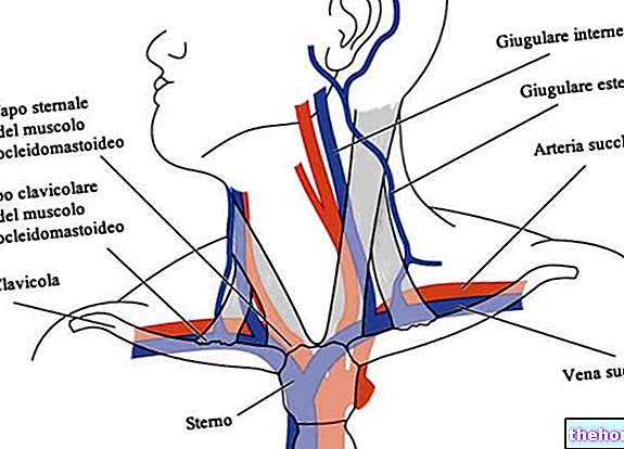
Located one to the right and one to the left of the neck, the external jugules drain blood into the subclavian veins, with which they are in communication. Clearly, the right external jugular drains blood into the right subclavian vein, while the left external jugular drains blood into the left subclavian vein.
The external jugularies collect much of the blood that has oxygenated the external part of the skull and the deeper tissues of the face.
Each external jugular arises from the conjunction between the posterior division of the retromandibular vein and the posterior auricular vein.
The external jugular veins each have two tributary veins (N.B: tributary means tributaries). In fact, for each external jugular vein, there are a posterior external jugular vein and an anterior jugular vein. The posterior external jugular vein collects the blood that has oxygenated the back of the neck; the anterior jugular vein, on the other hand, collects the blood that has oxygenated the larynx and all the tissues of the lower portion of the jaw.
As regards the course of the external jugular veins, these originate where the parotid gland resides, approximately at the height of the so-called angle of the mandible. From here, they descend perpendicularly along the neck, towards the clavicle. In the first part of this path, they are located on the posterior border of the sternocleidomastoid muscle; subsequently, they cross the latter obliquely, reaching the subclavian muscle. At the level of the subclavian muscle, they join the subclavian veins.
Each external jugular vein has two pairs of valves: a lower pair and an upper pair.
The pair of inferior valves resides at the point where an external jugular vein joins a subclavian vein; the pair of upper valves typically reside 4 centimeters higher than the collarbone. The jugular tract that is interposed between the two pairs of valves is called the sinus.
The aforementioned valves serve to facilitate the transport of blood towards the heart, but, contrary to what one might think, they do not prevent blood reflux; in other words, they do not prevent the blood from returning.
INTERNAL JUGULAR VEINS
Like the external jugularis, the two internal jugulars are also one on the right and one on the left, on the neck. Together with the corresponding subclavian veins in a more internal tract, compared to what happens for the external jugular, their task is to collect the blood that has oxygenated the brain, the meninges, the superficial tissues of the face and neck.
The confluence of the internal jugular veins into the subclavian veins occurs very close to where the latter become brachycephalic veins.
The left internal jugular vein is slightly smaller than the right internal jugular vein.
As regards the course of the internal jugular veins, these originate at the base of the skull, at the point where the so-called inferior petrosal sinus and the so-called sigmoid sinus join. According to some human anatomy texts, the starting point of the internal jugular veins would be at the level of the posterior compartment of the jugular hole (or foramen).
From the point of origin, the internal jugulars proceed along the neck in a vertical direction, occupying a lateral position, first, with respect to the internal carotid artery and, subsequently, to the common carotid artery.
In their vertical path along the neck, the internal jugules are also close to the vagus nerve.
Curiously, both shortly after their origin and shortly before their junction with the subclavian veins, the internal jugular veins have a swelling: the swelling present at the origin is called the superior bulb, while the swelling present almost at the level of the subclavian vein takes the lower bulb name.
Each internal jugular vein contains a pair of valves. Located about 2.5 centimeters higher than where the internal jugular veins end, these valves facilitate the transport of blood, but do not prevent blood reflux.
The jugular veins, the carotid arteries and the vagus nerve are comprised within the so-called carotid sheath. The carotid sheath is a thickening of the deep cervical fascia.
Function
The jugular cells contribute to the return to the heart of the blood that has recently oxygenated the various organs and tissues of the head.
This deoxygenated blood re-enters the right atrium of the heart, through the superior vena cava; once in the right atrium, the cardiac organ, thanks to its contractile capacity, introduces it first into the right ventricle and then into the lungs. The lungs represent the site where oxygen-poor blood is oxygenated.
Then, from the lungs, the blood returns to the heart, precisely in the left atrium; from the left atrium it passes to the left ventricle, which finally pumps it into the arterial system.
Clinic
The jugular muscles lack bone or cartilage protection, therefore they are extremely susceptible to damage and injury, which can result from, for example, a cut on the neck.
The lesions affecting the jugularis are responsible for a conspicuous loss of blood, as the volume of blood that passes through them is considerable.
JUGULAR VENOUS WRIST
The pressure of the blood circulating inside the jugular is a useful diagnostic parameter for identifying heart diseases such as heart failure, tricuspid valve stenosis, tricuspid regurgitation or cardiac tamponade.
The measurement of the blood pressure circulating in the jugular is called the jugular venous pulse.


