Generality
Otoscopy is an objective examination of the ear, which allows you to inspect the external auditory canal and the tympanic membrane; in this way, the doctor can identify the presence of foreign bodies and / or pathological conditions that can cause various disorders, such as, for example, ear pain, hearing loss or deafness.

Otoscope
As mentioned, otoscopy is performed through the use of an instrument called an otoscope.
There are several types of otoscope that can be used to conduct the aforementioned physical examination of the ear. However, the most common is, most likely, the battery-powered otoscope.
The battery-powered otoscope is an optical instrument whose shape resembles that of a hammer. It is composed of a base, or handle, which can be made of plastic or metal, and a head with which the "real exam.
The head of the otoscope has a cone shape; in it there are a luminous protrusion that directs the ray of light inside the ear and a magnifying glass made of optical glass that allows a magnified view (usually 3x) of the ear canal and eardrum.

At the final end of the cone that constitutes the head of the otoscope - before carrying out the examination - an ear speculum made of plastic and conical in shape is inserted. Usually, the speculum in question is disposable and must have an adequate size for the size of the patient's ear canal on which it is necessary to perform the examination (the ear specula used in children, therefore, will be smaller than those used for adults).

How Otoscopy Is Performed
Before proceeding with otoscopy, the doctor must first exert an upward and backward traction on the auricle, in order to "straighten" as much as possible the ear canal, which in itself tends to be curved and do not allow an "adequate view of" the external ear and the tympanic membrane.
After performing this simple maneuver, the doctor can insert the otoscope inside the patient's ear and proceed with the examination.
During otoscopy, usually, the doctor supports the index, or even the whole hand holding the otoscope on the patient's head, in order to be more stable and in order to avoid possible accidental lesions of the auditory canal due, in fact, to a possible instability in the grip of the instrument.
What is observed with the Otoscope
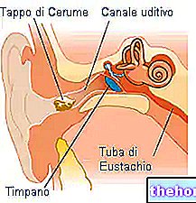
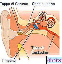
Through the use of the otoscope, the doctor (general or specialist) is able to inspect the external ear - therefore the auditory canal - and the tympanic membrane of the patients. More in detail, thanks to the use of this particular optical instrument, the doctor can detect the presence of:
- Abnormalities and / or malformations of the ear canal or tympanic membrane;
- Inflammation and / or pathologies of the external ear and / or of the middle ear (otitis, mycosis, etc.);
- Ear plugs and foreign bodies of various kinds.
Furthermore, the otoscope allows to examine the tympanic membrane, allowing the evaluation of parameters that characterize it, such as:
- Color;
- Position;
- Mobility;
- Brightness;
- Translucency.
In addition to this, thanks to the otoscopic examination it is also possible to identify the presence of any endotympanic effusions.
Micro-otoscopy
Micro-otoscopy is an objective examination of the ear exactly like otoscopy, but it differs from the latter because - instead of being performed with a normal battery-powered otoscope - it is performed through the use of a binocular microscope.
This instrument - characterized by the presence of a "coaxial illumination - allows to perform a micro-otoscopic examination, allowing a vision of the external ear and of the middle ear that is much larger (10x or 20x) than that obtained by means of the" use of the most classic battery-powered otoscope. Furthermore, thanks to the binocular microscope it is also possible to perform minor surgical interventions - on an outpatient basis, as well as in the operating room.
Operations and Surgical Interventions
Through otoscopy - in addition to performing an "analysis of the external and middle ear - it is also possible to perform some simple operations or small surgical interventions. The simplest operations can be performed in an outpatient setting by the" otolaryngologist, with the "use of" battery-operated otoscope and any other instruments; the more complex surgical interventions, on the other hand, are performed in the operating room and, generally, with the aid of the binocular microscope.
In any case, the main operations and small surgeries that can be performed otoscopically are:
- Removal of ear wax plugs and foreign bodies of various kinds (this operation can be performed with the use of the battery-powered otoscope by inserting a special instrument on its head to remove the body causing the obstruction of the ear canal);
- Removal of small polyps from the ear canal
- Tympanocentesis, ie the execution of a small incision on the tympanic membrane in order to evacuate or withdraw pus (mainly used in the case of purulent otitis);
- Transtympanic drainage (carried out mainly in cases of serous otitis).
Pneumatic otoscopy
Pneumatic otoscopy is an examination of the ear that is carried out in the pediatric setting mostly to diagnose acute otitis media, to evaluate its progression and healing and to evaluate the presence and possible persistence of endotympanic exudate.
This examination is carried out with the use of a particular instrument - the pneumatic otoscope - and allows to perform a more in-depth analysis of the tympanic membrane than that which can be carried out with simple otoscopy.
The pneumatic otoscope, in fact, is equipped with a connector on which a ball insufflator is positioned which - through the speculum - allows air to be blown into the ear to assess the mobility of the tympanic membrane.
Under normal conditions, the tympanic membrane moves freely under the influence of the jet of air that is pushed into the ear, while in pathological conditions there is an alteration in the movement of the same membrane. This alteration can manifest itself. , for example, as a reduction of the mobility of the membrane upon compression and release of the insufflator, or as a more accentuated mobility upon release of the insufflator rather than its compression.
Pneumatic otoscopy, therefore, is able to provide more detailed information than classic otoscopy, thus allowing to define more accurately the pathologies of the middle ear in pediatric age.

