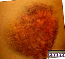
The ideal treatment for hematomas depends on the severity of the trauma suffered - or in any case on the severity of the underlying disease - and on the site involved.
in a tissue or organ, accumulated following the rupture of a blood vessel, whether this is a capillary or any other duct of the circulatory system. The blood, finding no way out, concentrates and accumulates in the compromised region.The hematoma, in addition to trauma and contusions, can be caused by several factors: impaired coagulation (thrombocytopenia, haemophilia), surgical wounds, leukemia and therapy with anticoagulant drugs (eg heparin, dicumarol, etc.).

Considering the heterogeneity of the triggering causes, it is understandable that there are many treatments for the hematoma.
to blue, then from purple to yellowish-green) is an important signal, a clue from which it is possible to understand that the hematoma is being reabsorbed.Nevertheless, it is possible to adopt some small measures to speed up the healing time of the hematoma, without taking drugs or undergoing surgical therapies.
A modest hematoma resorbs faster when treated with ice (cryotherapy): the application of an ice pack directly on the superficial hematoma promotes vasoconstriction, which limits the leakage of blood from the vessels injured by the contusion.
DO NOT apply ice directly on the hematoma: if you do not have a suitable ice bag, it is recommended to wrap some ice cubes (or other frozen products) in a cloth. Then, apply everything on the hematoma. This simple trick is indicated to reduce possible cold burns.
In addition to its extraordinary vasoconstrictive capacity, ice excellently exerts a "further therapeutic function: when applied to the hematoma, the cold pack creates a sort of anesthesia, thus acting as a mild anesthetic. The ice pack is placed directly on the superficial hematoma, and therefore acts as a mild anesthetic. left there for about ten minutes. Repeat the application several times during the day (about 2-3 applications every hour), for 2-3 days.
Only when the superficial hematoma is extensive and painful, complementary palliative care is recommended: in such circumstances, it is recommended to take NSAIDs, painkilling and anti-inflammatory drugs that temporarily mask the pain caused by the contusion. The application of anti-inflammatory ointments is also a good remedy to relieve pain.
In the case of a more severe sub-nail hematoma, the removal of the nail is conceivable.
If the hematoma is extensive and the contusion is violent, it is advisable to undergo a radiological test to rule out a possible fracture of the phalanx.
, but also and above all to remove the hematoma.
Under ultrasound guidance, the hematoma is emptied: this maneuver must be performed in a hospital setting, under aseptic conditions (complete sterility).
Cranial hematomas in general must be evacuated surgically: the removal of the mass consequently also reduces the pressure exerted by the hematoma on the brain. The hematoma is evacuated through a hole made directly in the skull (craniotomy).
Any concomitant infections must be treated with specific antibiotic therapy. For example, hematomas resulting from surgical wounds can favor the appearance of an infection, which requires immediate treatment to avoid the spread of pathogens to other districts.
Often, the tissue affected by the hematoma evolves into fibrosis, therefore there is an exaggerated increase in the fibrous connective component, to the detriment of the parenchymal cells. This situation, typical of muscle and subcutaneous hematomas, can originate calcifications, responsible for pain and thickening of the In this sense, the best cure for the healing of the hematoma is shock wave therapy: it is a useful therapeutic strategy to promote and increase local capillaryization and cellular metabolism, favoring the spontaneous repair process of the tissue. , hence the resorption of the hematoma.


