The orthoptic examination can also be useful in the presence of other ocular problems (eg traumatic accidents, work activities that are particularly tiring for the eyes, dyschromatopsies, etc.) and allows you to plan the most appropriate rehabilitation path for the patient.
binocular, verifying the degree of collaboration between the two eyes. This test is used, in particular, to confirm or exclude the presence of disorders of the neuromuscular system and the alterations that derive from these (such as strabismus, amblyopia, suppression mechanisms and ocular paralysis).
The orthoptic examination involves the evaluation of ocular motility, in the sense of:
- Three-dimensionality ("stereopsis");
- Convergence;
- Movements that allow the eyes a unitary vision;
- Accomodation.
The orthoptic examination is an assessment aimed at both children and adults.
Diagnostic scope
The orthoptic examination is especially useful to highlight the restrictions in the movement of the extraocular muscles in the various gaze positions, both for each single eye and when they work together (ocular motility examination).
The examination checks the convergence of the gaze and, in cases of strabismus, allows us to quantify the extent of the deviation, identify which muscle causes diplopia (double vision) and follow the evolution of the clinical picture over time.
The orthoptic examination also assesses sensitivity to color contrast and dyschromatopsia (difficulty in perceiving colors).
Orthotic treatment of visual rehabilitation
As regards the therapeutic field, the orthoptic examination plays an important role in monitoring amblyopia (a condition that produces the unilateral reduction of vision), since it follows the evolution of the improvement of vision by intervening, depending on the specific case, with Orthoptic exercises or any bandages. Orthoptics is also useful for defining rehabilitation pathways in patients suffering from neurological diseases or who have suffered head trauma, as well as providing support in case of postural alterations, dyslexia or learning disorders.
Orthoptic examination: when is it indicated?
The orthoptic examination is important for the diagnosis of various pathologies, which affect binocular vision, reduce motor skills (in particular, in driving or manual dexterity tasks that require speed and precision) and, in the child, can cause a delay in development (as in walking and talking).
In this regard, it should be noted that this "examination is part of the protocol dedicated to pediatric prevention:
- In the first 6-8 months of life, the orthoptic examination serves to exclude the presence of congenital pathologies or elevated vision defects that can cause significant damage to the vision, but if they are identified and treated early they are easier to manage.
- If everything goes smoothly, the next check should be carried out between two and three years. In this age group, the child is able to distinguish simple symbols and, if managed in a calm manner, collaborates with the ophthalmologist, for whom it will be easier to assess the presence of any vision defects, such as amblyopia.
- In preschool age (5-6 years), the ophthalmologist performs an even more accurate vision check than the previous one: the child, in fact, in addition to recognizing drawings and letters, can interact with the doctor by answering his questions. verify that the development of the visual system is proceeding correctly and that there are no difficulties in binocular cooperation such as to affect reading and writing.
In adulthood, the orthoptic examination is aimed at people suffering from general or specific pathologies of the visual system that induce symptoms such as diplopia, visual field alterations or postural defects.
(diplopia);
Following the orthoptic examination and other tests, the doctor will prescribe the most appropriate treatment for the ailment he has encountered. The orthoptic examination is also able to control the evolution of the pathologies already diagnosed.
, the evaluation begins with a check aimed at excluding the presence of limitations of the muscles responsible for moving the eyeballs, both for each single eye and in simultaneous vision.Subsequently, the ability to fix approaching objects (convergence) is checked and that there are no points in space where the vision is doubled.
During the examination, the doctor checks visual acuity, that is, he measures how clearly the patient is able to see; in general, the patient is asked to recognize some optotypes (graphic symbols, Albini's E, letters or numbers) arranged at a precise distance.
Once this first phase has been completed, the orthoptic examination involves the execution of specific tests that allow to deepen the clinical picture.
The main orthoptic tests
The most commonly used orthotic techniques include:
- Stereopsis: during the orthoptic visit, this test evaluates the sense of depth and three-dimensional vision, which can be defective if there is no correct synergy between the two eyes (as can happen, for example, for very different visual defects between one eye and the other).
- Convergence: it is an orthoptic test that evaluates the ability of the two eyes to make a harmonious and symmetrical movement when they are stimulated to converge, making an object that progressively approaches the tip of the nose to stare. This evaluation is very useful in subjects who use a VDU for a long time. As far as the convergence movement is concerned, the orthoptic examination can also verify the fusional amplitudes, i.e. the ability of the two eyes to collaborate in merging two distinct images into a single image and maintaining this uniqueness even when they are stimulated to converge or diverge.
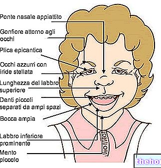
- Ocular motility examination (MOE): during the orthoptic examination, verifies the functionality of the muscles that move each eye, in the main gaze positions. This test allows to highlight a limited ocular motility, the possible misalignment of the eyes and nystagmus. The examination of ocular motility is used to identify the presence of hyper and / or hypofunctions of the extraocular muscles (such as, for example, the deficit of the external rectus muscle, which is involved in the paralysis of the 6th cranial nerve), anomalies of coordinated movement of the two eyes (e.g. convergence deficit), particular conformations of the facial massif such as to induce pseudo or real strabismus (e.g. epicanthus, orbital strabismus etc.).
- Test for the study of diplopia: this evaluation of the orthoptic examination verifies the occurrence of double vision (a "single image is perceived as double) and its relative nature (horizontal, vertical and oblique). The doctor therefore pays particular attention to the way in which the eyes concentrate and move together to focus a visual stimulus (alignment, convergence and focus). Any deficits found may suggest the presence of eye or eyelid injury, an orbital or retrobulbar disorder, etc.
- Cover test: serves to highlight the presence of strabismus, classifying them in manifest (always present) or latent (they emerge only in certain circumstances), as well as indicating in which direction the ocular deviation occurs (convergent, divergent, vertical or torsional). If combined with the use of a prism stick, the cover test allows to measure the power of the prismatic lens necessary to compensate for the deviation.
- Screen examination of Hess Lancaster and Gracis: in the presence of strabismus, this test of the orthoptic visit is used to quantify the degree of deviation and the state of the muscles affected by the problem. This examination is preliminary to surgery.
- Test for the evaluation of sensoriality: examines the binocular relationships and the retinal correspondence of the two eyes (ie how much the two images that are formed on the retina of the two eyes "match"). This test allows to detect the presence of sensory abnormalities of vision such as suppression (ie an eye is not used due to the poor quality of the image it provides to the brain). The fusional and accommodative performance is altered in case of strabismus and / or amblyopia.
How long does it last?
The duration of the orthoptic visit varies, but generally takes 15-20 minutes.
What does the report report?
The diagnostic conclusion is reported in the report of the orthoptic examination drawn up by the ophthalmologist.

