Radiology
A typical apparatus for traditional radiology is composed of several parts:
-
x-ray tube: it has the function of producing X-rays and directing their beam towards the desired target. It is housed in a casing (protective cap), which has the function of reducing the level of radiation and which can be oriented in different directions;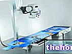
- current and electric potential generator: it supplies the electric current necessary to the X-ray tube for the production of the same;
- command table: allows the operator to set the patient's electrical current and exposure time according to the case;
- accessories: they are numerous, and vary according to the type of device and according to the type of investigation it is intended for;
- patient bed or table: it can be fixed in the horizontal position (trocoscope), or in the vertical one (otoscope), or it can be tilted from the vertical to the horizontal position, following various degrees of curvature (clinoscope). In some devices the bed can be suspended, that is suspended from the ceiling and, with a telescope mechanism, can be easily moved up or down;
- Support supports for the X-ray tube;
- Seriographers: they are devices that allow to perform numerous radiograms in series in a short or very short time (rapid serigraphs) and are intended for the study of moving organs or structures, whose dynamics must be evaluated. They are mainly used in the study of the digestive system and in angiography;
- Grids and anti-diffusion systems: they have the purpose of eliminating diffuse X radiation (not oriented on the anatomical site to be studied);
- Image or image intensifier: allows the radiological image to be displayed on a monitor.
How is the image formed in radiology?
Traditional radiology equipment can work in the radioscopy or in the radiography mode.
Radioscopy or fluoroscopy
There radioscopy or fluoroscopy exploits the property of X-rays to make some substances fluorescent, such as barium platinocyanide. If an X-ray beam hits a paper support on which a layer of fluorescent substance is deposited, this becomes luminous, because its molecules absorb X radiation, become excited and, in the subsequent return to the rest state, emit photons in the visible spectrum. (fluorescence). The fluorescent layer therefore transmits light in proportion to the "intensity of X radiation that hits it. If a radiopaque body (like the human one) is interposed between the X-ray source (X-ray tube) and the fluorescent layer (X-ray screen), the" light effect does not occur where the radiation absorbed (stopped) by the radiopaque body does not reach the screen. On the latter, therefore, the image of the body appears in positive, ie dark. In the case of the human body, this effect is complex because the body is made up of various substances, very different from each other. In fact, the "absorption of X rays (ie the ability to prevent their passage) varies according to the atomic number of the substances that make up the body and, for the same atomic number, the thickness of the body. Some parts of the" organism, therefore, due to their high atomic number and their consistent thickness, they almost completely retain radiation; others retain them only partially; others, finally, allow them to pass almost completely. The former appear dark on the radioscopic screen, the latter are gray, with different degrees of intensity, while the thirds appear clear. For example, if the chest of a man is interposed between the X-ray tube and the radioscopic screen, with the emission of X-rays the bones (ribs) and the mediastinum are observed on the dark screen, the soft parts (muscles, vessels, etc.) are gray, the lungs are clear. The whole of all these components, which have different shades of brightness, constitutes the radioscopic image of the thorax.
Radioscopy is used in all investigations where direct vision of the examined object is required. For example, in the study of the heart or the great central vessels, in which it is called angiography, a catheter is introduced into a vein or a " peripheral artery. This catheter is followed with radioscopic control in its progression to the desired point.
Other articles on "Radiology and radioscopy"
- Radiography and X-rays
- X-ray


