Generality
MOC, or Computerized Bone Mineralometry, is a diagnostic test that measures the levels of calcium and other minerals in the bones of the human skeleton.

There are several types of MOCs. The most common type is MOC DEXA; the MOC DEXA measures the BDM using an X-ray instrument.
MOC is mainly indicated for: the diagnosis of osteopenia or osteoporosis, the evaluation of the effect of treatments for osteopenia or osteoporosis and the monitoring of all those conditions that can cause osteopenia or secondary osteoporosis.
MOC does not require special preparation, lasts between 20 and 30 minutes and does not require any hospitalization.
Typically, the results of the MOC are ready 2-3 days after the examination is performed.
What is the MOC?
MOC is a diagnostic test for measuring the levels of calcium and other minerals in the bones of the human skeleton.
Through the MOC, doctors estimate the so-called bone mineral density (BDM, from English Bone Mass Density).
Bone mineral density is a measure of the amount of minerals (bone mineral mass) contained in one cubic centimeter of bone (volume).
WHAT DOES MINERAL AND BONE DENSITY INDICATE?
Bone mineral density is an indicator of bone resistance to fractures: lower than normal BDM values are indicative of a certain bone fragility and a certain susceptibility, on the part of the skeleton, to suffering fractures.
WHAT DOES MOC MEAN?
The acronym MOC is the acronym for Computerized Bone Mineralometry.
Types
There are several types of MOCs. What distinguishes each type of MOC is the technique of measuring bone mineral density.
The most common type, and the one to which the next chapters of this article will refer only with the term of MOC, is the so-called MOC DEXA. Also known as Double Energy X-ray Absorbimetry (in English Dual-Energy X-ray Absorptiometry), the MOC DEXA provides, for the quantification of bone mineral density, the use of X-rays at two different energy levels.
Among the types less used, compared to the MOC DEXA, we note:
- The SPA, or the "Single Photon Beam Absorbimetry. It involves the use of a radioactive substance. It is a measurement technique that was more in vogue in the past.
- The DPA, that is the Double Photonic Beam Absorbimetry. Like the previous SPA, foresees the use of a radioactive substance. It is an increasingly less practiced measurement technique.
- Quantitative Computed Tomography. Also known as MOC QTC, it works similar to regular Computed Axial Tomography.
- Quantitative bone ultrasound, also known as MOC QUS, involves the use of ultrasounds, which are not harmful to humans at all.
Its diagnostic power, however, is lower than that of previous types of MOC.
Uses
MOC is a test indicated for various purposes, including:
- The diagnosis of conditions such as osteoporosis and osteopenia, which are characterized by a reduction, compared to the levels considered normal, in bone mineral density;
- Evaluation of how and if medications for the aforementioned conditions are working. Knowing how effective a certain drug therapy is is useful for the doctor to understand whether or not it is necessary to make a change;
- Monitoring of the skeletal effects of prolonged intakes of corticosteroid drugs (e.g. prednisone). Corticosteroids are powerful anti-inflammatories, the use of which for long periods of time can have several side effects, including osteopenia or osteoporosis.
- The estimate of how much an individual is at risk of skeletal fractures;
- Monitoring of conditions which, among the various symptoms caused, also determine a lowering of bone mineral density (osteopenia or secondary osteoporosis). The conditions in question include: multiple myeloma, Cushing's syndrome, hyperthyroidism, hyperparathyroidism, premature menopause, vitamin D deficiency and alcoholism.
OSTEOPOROSIS AND OSTEOPENIA
Osteoporosis is a common systemic disease of the skeleton, which causes a strong weakening of the bones. This weakening originates from the deterioration of the microarchitecture of the bone tissue and the consequent reduction of the bone mineral mass. Due to the aforementioned bone weakening, the bones of people with osteoporosis are more fragile and prone to fractures.
Osteopenia is a very similar condition to osteoporosis; to distinguish it from the latter are the lower degree of reduction in bone mineral density and the consequent lower risk of skeletal fractures. In other words, osteopenia is a mild form of osteoporosis.
Osteopenia and osteoporosis are two typical conditions of old age: in the female population, it is particularly common from the age of 65 onwards, in the male population, on the other hand, it is particularly common from the age of 70 onwards.
The tendency of the elderly to develop osteopenia and osteoporosis is why doctors recommend that women aged 65 and over with a past history of fractures and men aged 70 and over with a past history of fractures do. MOC every two years.
- Familial predisposition to decrease in bone mineral density
- The reduction of estrogen levels in women and the reduction of testosterone in men
- Alcohol abuse
- Cigarette smoke
- Poor calcium intake in the diet
- Anorexia nervosa
- Excessive thinness
- The sedentary lifestyle
- The advanced age
Preparation
The MOC does not require any particular preparation.
In fact, the only preparatory measure, which patients must comply with, concerns the clothing to wear on the day of the examination and consists in avoiding any clothing or accessories (eg necklaces, earrings, etc.) with metal parts.
Procedure
Typically, the places where MOC takes place are the radiology department of hospitals or hospital clinics specializing in radiology and diagnostic imaging techniques.
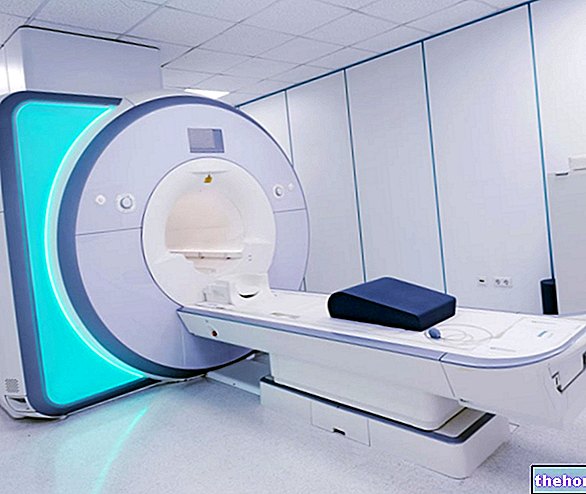
The first part of the procedure consists in the accommodation and positioning of the patient, on the table combined with the instrument for the emission of X-rays, necessary for measuring the bone mineral density.
For the success of the examination, the patient must maintain the position that the radiologist or radiologist has imposed.
Unless otherwise indicated, undressing is not envisaged.
Only once the positioning is finished, can the second part of the procedure begin, which consists of exposing the patient to X-rays.
After the exposure, the test can be considered concluded. The patient can therefore get up from the table on which he was lying and immediately return home.
WHAT HAPPENS DURING X-RAY EXPOSURE?
Without too many details, in hitting the patient's body, the x-rays are absorbed, in part, by the soft tissues, and in part, by the bone tissues.
While soft tissue uptake is useless for diagnostic purposes, bone uptake is what is needed for quantification of bone mineral density.
This absorption, in fact, varies in relation to the bone mineral mass of the subject examined.
IN WHICH BONES DOES THE MEASUREMENT OF BONE MINERAL DENSITY TAKE PLACE?
On the occasion of the MOC, the bones from which doctors generally obtain the bone mineral density values of an individual are: the vertebrae of the spinal column, the bony portions that make up the two joints of the hip, the most important thoracic bones, the " ulna and radius of the forearm, the phalanges of the toes or hands and the heel bone.

