Generality
Renal scintigraphy is a diagnostic test of nuclear medicine, which allows to study in detail the anatomy and function of the kidneys, detecting any abnormalities.

The preparatory rules for the exam are few and simple to follow; among these, fasting does not appear.
Depending on what the research topic is, a renal scintigraphy can last from 30 to 120 minutes.
The risks are minimal.
Thanks to renal scintigraphy, doctors are able to recognize and evaluate conditions such as: kidney failure, kidney cysts or tumors, kidney inflammation or infections, blockages of the kidney arteries, changes in the flow of urine inside kidney, kidney transplant complications, renovascular hypertension, hydronephrosis, etc.
Brief review of the kidneys
The urinary system or excretory system is the set of organs and anatomical structures responsible for the elimination of urine.
The main organs of the urinary system are the kidneys.
Two in number, the kidneys reside in the abdominal cavity, on the sides of the last thoracic vertebrae and the first lumbar vertebrae; they are symmetrical and have a shape that is very reminiscent of that of a bean.
The most important functions of the kidneys are:
- Filter waste substances, harmful substances and foreign substances present in the blood and convert them into urine.
- Regulate the hydro-saline balance of the blood.
- Regulate the acid-base balance of the blood.
- Produce the glycoprotein erythropoietin.
The anatomy of the kidneys is quite complex: the image below shows the main anatomical elements of a generic human kidney.

What is renal scintigraphy?
Renal scintigraphy is a diagnostic test of nuclear medicine, which, through the venous injection of radiopharmaceuticals, allows to accurately analyze the anatomy and function of the kidneys, and to determine if the latter are "working" properly or no.
In addition to radiopharmaceuticals, the execution of a renal scintigraphy involves the use of a special equipment - the so-called gamma camera - capable of reproducing in images the distribution of the radiopharmaceutical inside the organism.
The gamma camera is connected to a computer that is used to adjust its operation.
A nuclear medicine specialist, also called a nuclear doctor, performs the renal scintigraphy and above all to interpret the results.
Unlike the CT or radiographs, renal scintigraphy does not emit radiation, but detects the radiation emitted by the patient once the radiopharmaceutical is injected.
What is nuclear medicine?
Nuclear medicine is that branch of medicine that is based on the use of radioactive substances (the so-called radiopharmaceuticals) for diagnostic and therapeutic purposes.
A radiopharmaceutical is an injectable medicine that contains radionuclides, which are radioactive isotopes.
Once injected, radiopharmaceuticals can interact specifically with a specific biological tissue (like a normal drug) and, by virtue of their radioactive properties, can be monitored by means of a "special radioactivity detection instrument (gamma camera). In this way , this complex instrumentation provides very clear images of how, over time, the radiopharmaceutical is distributed inside the organism.
Scintigraphy is a diagnostic test of nuclear medicine, based on the detection of the radiations emitted by an organism after the administration, in the latter, of radiopharmaceuticals. These radiations, suitably processed by an instrument ad hoc, allow you to investigate the location, shape, size and function of various organs, including the heart, thyroid, bones, brain, liver, kidneys and lungs.
In the light of what has just been stated, it is possible to understand why radionuclides are also identified with the word "tracers".
Please note: the distribution in a certain tissue of the radioactive isotopes or their interaction with a given organ depends exclusively on the drug to which the aforementioned radioactive isotopes are bound. Therefore, the choice of the pharmacological preparation is of fundamental importance for the correct execution of the examination. For example, for a thyroid analysis it is necessary to resort to the use of a drug that diffuses specifically in this organ of the body; the same applies to the heart, kidneys etc.
Indications
Renal scintigraphy allows not only to identify the causes of a possible malfunction of the kidneys, but also to clarify whether a certain treatment, applied for the treatment of some kidney disease, is having an effect or not.
Going into more detail, through renal scintigraphy doctors are able to recognize and evaluate:
- A reduction in the blood supply to the kidneys;
- A state of renovascular hypertension, ie high blood pressure due to a narrowing of the arteries that carry blood to the kidneys;
- Kidney cysts or tumors
- A kidney abscess
- The results of renal medical treatment;
- The outcome and complications of a kidney transplant;
- L "acute or chronic renal failure;
- A "kidney infection (pyelonephritis);
- Injuries to the kidneys or related structures (ex: ureters);
- A state of obstructive uropathy;
- L "hydronephrosis;
- A glomerulonephritis.
Types of renal scintigraphy
Renal scintigraphy includes 4 different techniques for studying the function of the kidneys. The techniques in question are:
- Static or cortical renal scintigraphy. It provides information on the functioning of the cortical portion of the kidneys.
With this technique, the collection of images with the gamma camera can only take place after about 2 hours have elapsed from the administration of the radiopharmaceutical. - The perfusional-functional renal scintigraphy. It has various purposes: it shows the blood flow inside the kidneys, it detects any narrowing of the renal arteries and, finally, it helps to understand how the kidneys are working.
With this technique, imaging with the gamma camera can take place immediately after the injection of the radiopharmaceutical; typically, doctors take a picture of the kidney situation every 20-30 minutes. - [Renal diuretic scintigraphy. Identify any blockages or obstructions in urinary flow within the kidneys.
This technique involves observing the flow of urine in the kidneys both before and after the patient takes a diuretic, ie a stimulant of diuresis. - Sequential renal scintigraphy with ACE inhibitors. Thanks to the use of a drug against high blood pressure (ACE inhibitor), this technique allows us to understand if the hypertension present in an individual is due to a narrowing of the renal arteries or not.
For a correct and profitable execution of sequential renal scintigraphy with ACE inhibitors, it is essential to observe the kidneys both before and after the administration of the antihypertensive drug.
Preparation
Renal scintigraphy does not require special preparation. In fact, the only preparatory rules to which the patient must comply are:
- Communicate to the doctor who prescribes the renal scintigraphy any pharmacological intake taking place at that time, since there are medicines, some even very widespread, capable of polluting the results of the diagnostic test in question. Among the medicines that, if used, can alter the "outcome of renal scintigraphy, in particular: ACE inhibitors (for the treatment of high blood pressure and some heart diseases), beta-blockers (also for the treatment of hypertension and heart diseases), diuretics and Non-Steroidal Anti-Inflammatory Drugs or NSAIDs (including aspirin and ibuprofen).
Once the patient has communicated the drugs he is taking, the doctor will decide what to do: for example, a possible solution is the request to temporarily interrupt the drug intake in progress and resume it only after performing the renal scintigraphy. - Always report to the doctor who prescribes the renal scintigraphy any allergies and pathological conditions in progress or in the recent past.
In the event that the patient is a woman, also communicate a "possible state of pregnancy or presumed such. - In the hours shortly before the renal scintigraphy, drink plenty of water (at least 1 liter of water) so that, at the time of the examination, the bladder is full.
- Go to the examination preferably without jewelry or clothing with metal parts, as these could interfere with the success of the renal scintigraphy.
Is there a fast?
Unless otherwise indicated by the doctor, fasting is not part of the preparatory rules. Therefore, the patient can eat as "is his habit."
Procedure
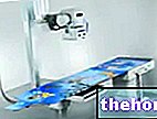
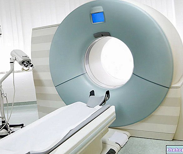
First, the patient must deprive himself of all objects and clothing that could interfere with the success of the renal scintigraphy; after which, he must lie down on a suitable sliding bed, which serves to position him between the plates of the gamma camera (see figure).

To position himself correctly on the couch, the patient can count on the help of a member of the medical staff, usually a technician from the nuclear medicine department or a general nurse.
At this point, with the patient lying down and before the "introduction" of the latter into the gamma camera, the nuclear doctor intervenes, who, thanks also to the collaboration of a professional nurse, carries out the injection of the radiopharmaceutical necessary for renal scintigraphy.
The injection of the radiopharmaceutical usually takes place in a vein in the arm or hand; the radiopharmaceutical takes a few minutes to distribute itself through the blood to the organs under investigation, namely the kidneys.
After the time necessary for the radiopharmaceutical to be distributed in the kidneys, the patient can finally be introduced between the plates of the gamma camera, and the detection of radioactivity can begin.
It should be noted that, at this point of the procedure, renal scintigraphy can vary from circumstance to circumstance, depending on its final purpose: for example, if the search for possible obstructions to the flow of urine in the kidneys is foreseen, the examination requires , at a certain time, the administration of a diuretic; if instead the object of diagnostic research is the narrowing of the renal arteries, the test involves, at a certain moment, the use of an ACE inhibitor; and so on (see what is stated regarding the types of renal scintigraphy).
While collecting images via the gamma camera, the patient must remain motionless; its movements, in fact, could alter the outcome of the diagnostic examination and the precision of the images.
Once the images necessary for a detailed assessment of the renal situation have been acquired, the nuclear doctor declares the renal scintigraphy completed and allows the patient to be extracted from the gamma camera, which a ward technician or general nurse takes care of.
Once dressed, the patient can return home immediately and resume his normal daily activities, unless otherwise indicated by the doctor.
During a renal scintigraphy, the most metabolically active kidney areas accumulate a lot of radiopharmaceutical, while the less active ones accumulate in small quantities. The areas with the highest concentration of radiopharmaceuticals are called "hot spots", while those with a reduced concentration of radiopharmaceuticals are called "cold spots".
How long does a kidney scan last?
A kidney scan can last anywhere from 30 minutes to 2 hours.
Its length depends on the purpose (for example, taking drugs during the procedure lengthens the time) and on the number of images that the nuclear doctor intends to collect in order to make an accurate diagnosis.
Recommendations to the patient immediately after the examination
Immediately after the examination, the doctor limits himself to recommending the patient to drink plenty of water, to favor the natural elimination of the radiopharmaceutical.
If the patient follows this recommendation, he eliminates the radiopharmaceutical from his body within 24 hours.
What sensations does the patient experience during the procedure?
The patient may experience some sort of discomfort when inserting the needle, due to the injection of the radiopharmaceutical.
Also, after the injection, you may experience a strange metallic taste.
Patients who struggle to remain still may feel uncomfortable after a short time; however, immobility is a fundamental condition for the successful outcome of the examination.
When might the patient have to wait before going home?
The patient may have to wait before returning home, in the event that unclear images emerge from the renal scintigraphy, which require a repetition of the examination or part of it.
Risks
Renal scintigraphy is a procedure that, in the trusted opinion of doctors and experts, is safe.
However, remember that:
- It involves exposing the patient (especially kidney and bladder) to a dose of harmful radiation.
Extent of the risk? Fortunately, the radiation dose is minimal. - In some people, the radionuclides (or radioactive isotopes) that make up radiopharmaceuticals could give rise to unpleasant allergic reactions (anaphylactic reactions).
Extent of the risk? Allergic reactions after renal scintigraphy are a very rare occurrence, affecting very few patients. - Given the relatively recent birth of nuclear medicine, the long-term effects it can have on human health are unknown.
- The needle used to inject the radiopharmaceutical may be responsible for a small wound and redness where it is inserted.
Extent of the risk? The wound and redness resolve within 24-48 hours.
Contraindications
They certainly represent a contraindication to renal scintigraphy: pregnancy, breastfeeding and failure to interrupt pharmacological treatments based on ACE inhibitors.
Results
Depending on the severity of the patient's condition, renal scan results may be available immediately or after a few days.
A renal scintigraphy with a clinically relevant outcome (therefore with abnormal results) occurs in the presence of conditions, such as: renal failure (both acute and chronic), cysts or renal tumors (NB: renal scintigraphy does not distinguish the two circumstances ), kidney inflammation or infections, blockages of the renal arteries, changes in the flow of urine inside the kidneys, complications of a kidney transplant, renal hypertension, hydronephrosis, etc.
Benefits
Renal scintigraphy provides information that is often impossible to obtain with other diagnostic procedures.

