-a-cosa-serve.jpg)
The fundus examination allows to detect the presence and monitor the progression of some pathologies, such as retinal detachment, age-related macular degeneration and other forms of maculopathy. symptoms such as lacunar defects (scotomas, phosphenes) and loss of half of the visual field (hemianopia), floaters (the so-called flying flies).
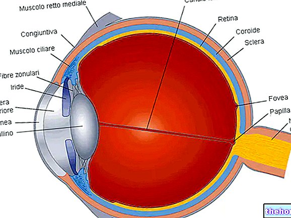
, that is the vitreous body, the retina (in particular, the macula, the central retinal area) and the papilla of the optic nerve.The fundus examination is sometimes referred to as ophthalmoscopy, referring to the instrument used by the ophthalmologist to study the back of the eye: the ophthalmoscope.
of gelatinous consistency, colorless and transparent, which occupies the cavity of the eyeball between the posterior surface of the lens and the retina (vitreous chamber). This mass helps to maintain the shape of the eye (fills the bulb), promotes the diffusion of nutrients and protects against microtraumas coming from the outside (absorbs shocks). Furthermore, being transparent, the vitreous body represents a means of refraction and, as such, allows the unobstructed transmission of light up to the retina (dioptric function). With the examination of the fundus, it is possible to verify the presence of its degeneration or any vascular anomalies.

