Observable in one in every 68,000-88,000 newborns, Apert syndrome is due to the specific mutation of the FGFR2 gene, which has the task of regulating the fusion of the cranial sutures and the development of the fingers and toes.
For the diagnosis of Apert syndrome, a physical examination, anamnesis, a radiological evaluation of the skull and fingers and toes, and, finally, a genetic test are fundamental.
Currently, those suffering from Apert syndrome can only count on symptomatic treatments, that is, they relieve the symptoms and avoid the most serious complications.
Brief review of the cranial sutures and their fusion
The cranial sutures are the fibrous joints, which serve to fuse together the bones of the cranial vault (ie the frontal, temporal, parietal and occipital bones).
Under normal conditions, the process of fusion of the cranial sutures takes place in the postnatal period, starting at 1-2 years of age, for some joint elements, and ending at the age of 20, for others. This long and cadenced process of fusion allows the brain to grow and develop adequately.
Apert syndrome, however, owes its notoriety not only to its association with craniostenosis, but also to the fact that it is related to a certain degree of syndactyly, that is the congenital anomaly characterized by the fusion of one or more fingers or fingers. feet.
The possibility of causing craniostenosis and syndactyly at the same time makes Apert's syndrome an example of acrocephalosyndactyly; in medicine, an "acrocephalosyndactyly is a genetic condition that combines specific malformations of the skull (" acrocephalus "means" head to tip ") with the fusion of one or more fingers or toes.
What are the consequences of early cranial suture fusion?
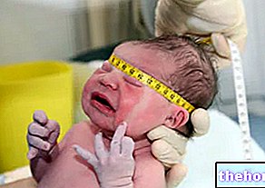
If, as in the case of Apert syndrome and other related diseases, the fusion of the cranial sutures occurs during prenatal, perinatal (*) or very early childhood, brain organs such as the brain, cerebellum and brainstem, and sense as the eyes undergo alterations in both growth and shape.
* N.B: "perinatal life" indicates the period between the 27th week of gestation and the first 28 days following childbirth.
Epidemiology: How Common is Apert Syndrome?
According to statistics, one in every 65,000-88,000 individuals is born with Apert syndrome.
Did you know that ...
The genetic diseases that, like Apert syndrome, cause craniosynostosis are about 150.
Among these, in addition to the Apert syndrome, the Crouzon syndrome, the Pfeiffer syndrome and the Saethre-Chotzen syndrome stand out in importance.
Curiosity
The acquired mutation that causes Apert syndrome is an example of a "mutation de novo", that is, of" new mutation completely devoid of a hereditary nature ".
What causes the gene mutation associated with Apert syndrome?
Premise: the genes present on human chromosomes are DNA sequences that have the task of producing fundamental proteins in biological processes essential to life, including cell growth and replication.
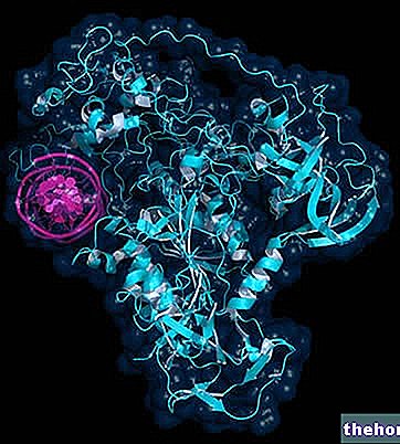
When it is free of mutations (therefore in a healthy person), the FGFR2 gene involved in Apert syndrome produces in the right quantities a receptor protein, called the Fibroblast Growth Factor Receptor 2, which is essential to mark the timing of fusion of the cranial sutures and to monitor the separation of the fingers and toes (in other words, it signals when it is the appropriate time for the fusion of the cranial sutures and regulates the formation of the fingers and toes).
On the other hand, when it undergoes the mutation observed in the presence of Apert syndrome, the FGFR2 gene is hyperactive and produces the aforementioned receptor protein in such massive quantities, that the timing relating to the fusion of the cranial sutures is altered (it is faster) and the processes of separation of the fingers and toes do not happen correctly.
Who is most at risk?
Regarding the acquired cases of Apert syndrome, the factors that induce the mutation of the FGFR2 gene after conception are not absolutely clear at the moment.
Research into this aspect is still ongoing.
Apert syndrome is an autosomal dominant disease
To understand...
Each human gene is present in two copies, called alleles, one of maternal origin and one of paternal origin.
Apert syndrome has all the characteristics of an autosomal dominant disease.
A genetic disease is autosomal dominant when the mutation of a single copy of the gene that causes it is sufficient to manifest itself.
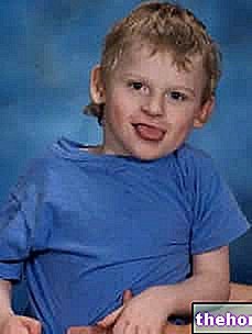
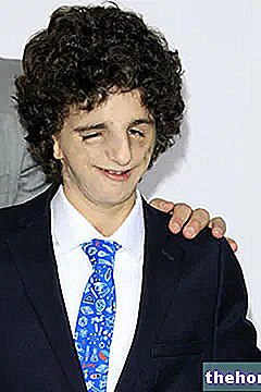
- Flat or concave face (due to deficient growth of the central bones of the face)
- Puffy, bulging, and wide-open eyes shallow eye sockets and abnormally spaced eyes (hypertelorism of the eye sockets);
- Beaked nose;
- Underdeveloped jaw, combined with prognathism;
- Crowded teeth (due to underdeveloped jaw)
- Ears lower than normal.
Syndactyly
In Apert syndrome carriers, syndactyly is seen in the hands, almost always, and in the feet, less frequently than in the hands.

The typical characteristics of syndactyly in the hands of an individual with Apert syndrome are 4:
- Presence of a short thumb with radial deviation (ie oriented abnormally towards the radius, one of the two bones of the forearm);
- Complex syndactyly between the index finger, middle finger and ring finger. By complex syndactyly, doctors mean an abnormal fusion of the fingers that affects not only the soft tissues (the skin), but also the bone tissues (the phalanges);
- Symphalangism. It is the medical term that indicates the anomalous fusion of the interphalangeal joints of the fingers (the interphalangeal joints are the joint elements present between the phalanx and the phalanx);
- Simple syndactyly between the fourth and fifth toes (ie between the ring and little fingers). With simple syndactyly, experts refer to an abnormal fusion of the fingers that affects only the soft tissues (the skin).
SEVERITY OF SYNDROME IN OPEN SYNDROME: THE 3 TYPES
Based on the severity of the thumb malformation (first of four characteristics), Apert syndrome experts distinguish 3 types of syndactyly of increasing severity:
- Type I (the least severe) coincides with a "minimal anomaly affecting the thumb, which remains totally independent of the index".
Other anomalies: index, middle and ring fingers are fused together through a complex syndactyly and present symphalangism affecting the distal interphalangeal joints; c "is simple and incomplete syndactyly between ring and little fingers (incomplete syndactyly means that the fusion between two fingers is partial).
Other information: is the most common type. - Type II (intermediate severity) is characterized by a more marked radial deviation of the thumb, compared to the previous case, and by a principle of syndactyly between the thumb and the index finger (c "is an incomplete syndactyly between thumb and index finger).
Other anomalies: index, middle and ring fingers are the protagonists of a complex syndactyly combined with distal symphalangism; between ring finger and little finger c "is a simple and incomplete syndactyly.
Other information: it is the second most common type. - Type III (the most severe) is characterized by the presence of a thumb joined in full to the index, not only at the level of the soft tissues but also at the level of the bone tissues.
Other anomalies: all the fingers are fused together, so much so that it is almost impossible to recognize them; c "is a" single nail; if between the first 4 fingers the syndactyly is complex, between the ring finger and little finger it is (as for the other types) simple and incomplete.
Other information: it is the rarest type.
Other possible symptoms and signs of Apert syndrome
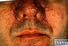
In some cases, in addition to being associated with craniosynostosis and syndactyly, Apert syndrome is related to the presence of: polydactyly (ie the presence of an extra finger in the hands or feet), hearing loss, recurrent ear and sinuses, hyperhidrosis, oily skin, severe acne, no hair on the eyebrows, fusion of the cervical vertebrae, obstructive sleep apnea syndrome and / or cleft palate.
Complications
The complications of Apert syndrome are, above all, the serious consequences that craniosynostosis can have on the development of the brain and intellectual abilities, and on the functional capabilities of the hands subject to syndactyly.
When is it possible to detect Apert syndrome?
Typically, cranial and digital abnormalities due to Apert syndrome are evident at birth, so diagnosis and treatment planning are immediate.
to the head (X-rays of the head, CT scan of the head and / or MRI of the head) and of the hands and possibly feet; finally, it ends with a genetic test.
Physical examination and medical history
Physical examination and anamnesis essentially consist in an accurate examination of the symptoms exhibited by the patient.
In a context of Apert syndrome, it is in these moments of the diagnostic process that the doctor finds craniosynostosis and syndactyly, and their precise characteristics.
Radiological examinations of the head and fingers and toes
In the context of Apert syndrome:
- Radiological examinations of the head are used by the doctor to confirm the presence of an early fusion of the coronal sutures (coronal craniosostenosis or brachycephaly); moreover, they allow him to estimate the severity of the cranial-brain anomalies in progress.
- On the other hand, radiological examinations of the fingers and toes are essential not so much for the confirmation of syndactyly (for this the visual examination is sufficient), but rather to know in detail the characteristics of interdigital fusions (type of syndactyly present, level of fusion etc.).
Genetic test
It is the DNA analysis aimed at detecting mutations in critical genes.
In the context of Apert syndrome, it represents the confirmatory diagnostic test, as it brings to light the FGFR2 mutation characteristic of the genetic disease in question.
THE SURGICAL CARE OF BRACHYCEPHALIA
For the carrier of Apert syndrome, surgical treatment of brachycephaly includes:
- A first intervention at a young age (within the year of life), aimed at separating the coronal fushes sutures earlier than expected. If this intervention is successful, the brain enjoys the right space for growth and there is a reduced risk of intellectual problems .
- A second intervention between the ages of 4 and 12, aimed at giving a normal appearance to the face, which (as the reader will recall) is flat if not concave.
The operation in question involves the incision of some bones of the face and their repositioning according to an arrangement that at least partially reflects normality. - A third eventual intervention in the years of childhood, with the aim of eliminating or at least reducing ocular hypertelorism.
THE SURGICAL CARE OF SYNDACY
Surgical treatment of syndactyly varies according to the characteristics of the interdigital fusion (so it depends on the type).

This means that the intervention valid for an individual with Apert syndrome may not be as valid for another individual with the same genetic disease (it is valid only if the type of syndactyly present is the same).
Having clarified this basic aspect, the goal of each type of existing surgical approach is the same and consists in releasing the fused fingers, in order to guarantee a certain functionality to the hands.
Generally, the treatment of syndactyly involves two stages:
- 1 step: "free" the first interdigital space (space between thumb and forefinger) and the fourth interdigital space (space between ring finger and little finger);
- 2 step: "free" the second and third interdigital space (space between index and middle finger, and space between middle and ring finger).

