What is the Ocular Orbit
The eye socket is a conical-pyramidal hexocranial cavity that contains and protects the eye.
Many bones of the skull (neurocranium) and the facial massif (splanchnocranium) articulate to form the orbital complex. This bone space therefore represents an anatomical crossroads in which blood vessels, nerve fibers, muscles, lacrimal glands and other annexes essential for the proper functioning of the organ of vision converge.
Pathologies of the orbit can be vascular, malformative, secondary to thyroid disease (Graves' disease), infectious, traumatic, inflammatory or neoplastic.
Anatomy
Eyeballs and relationship with the orbits
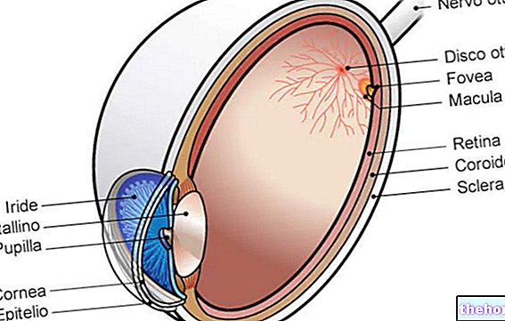
The eyes are two spheroidal formations with an average diameter of 24 mm (to be clear, they are slightly smaller than a ping pong ball) and a weight of 8 g. Each bulb occupies the orbital cavity together with the extrinsic muscles of the eye, the lacrimal gland, the cranial nerves and blood vessels also directed to the adjacent portions of the orbit and to the face. A fat pad (called the adipose body of the "orbit) has filling and isolation functions.

Orbital cavities
The orbits are two cavity formations placed on the sides of the midline of the face, below the forehead, made up of bones of the face and skull, in close correlation with each other.
From the morphological point of view, the orbit is comparable to a quadrangular pyramid, inverted backwards (with the apex back and the base forward), in which it is possible to distinguish:
- Base: represents the external opening of the orbit. The following take part in its formation: frontal bone and sphenoid (upper margin); maxillary, palatine and zygomatic (lower margin); ethmoid, lacrimal and frontal bone (medial margin); zygomatic and sphenoid (lateral margin).
- Upper wall: constitutes the vault or roof of the orbit; it is bounded by the lower face of the frontal bone and the lower face of the small wing of the sphenoid.
- Lateral wall: it is formed by the orbital process of the zygomatic bone and by the anterior portion of the large wing of the sphenoid.
- Medial wall: it is a sagittal bone plane formed by maxillary and lacrimal bone, papyrus lamina of the ethmoid and lateral face of the body of the sphenoid.
- Lower wall: represents the floor of the orbit and borders the upper face of the maxillary body, the upper face of the orbital process of the zygomatic bone and the orbital process of the palatine bone. Due to its thin thickness, the lower wall is the portion more frequently involved in orbital trauma.
- Apex: the posterior vertex of the orbit corresponds to the optic hole, crossed by veins, arteries and the optic nerve; this structure ensures communication between the eye and the middle cranial fossa.
Orifices and openings
The relationship between the bones of the orbital complex, although very close, is not absolute; the orbital walls have, in fact, holes and cracks that put this space in communication with the adjacent structures. These openings cross, in particular, the posterior end of the orbital cavity, at the apex (optic canal) or are located between the sphenoid and the maxillary bone (superior and inferior orbital fissure).
Functions
The orbits perform a protective and restraining function for the ocular structures, as they surround each bulb. In addition, they connect the eyeball to the rest of the organism.
Illnesses
Orbital disorders are generally inflammatory, traumatic, autoimmune or neoplastic in nature. Infiltrative ophthalmopathy caused by Graves' disease is the most frequent cause of orbital disease. Fractures of the orbit, on the other hand, account for approximately 40% of all craniofacial trauma.
The most common symptoms determined by the involvement of the orbit in the various pathological processes are represented by pain in eye movements, visual field changes, double vision and decreased vision. Orbital pathologies can also determine an "alteration of the normal positioning of the bulb. ocular in the orbit. The following can be observed: exophthalmos (bulbar protrusion), deviation (dislocation of the eye) and enophthalmos (hollowing).
In any case, a thorough eye examination is recommended and often, to confirm the diagnosis, investigations such as orbital ultrasound (studies the orbital contents), computed tomography (visualizes the orbital bone walls), nuclear magnetic resonance imaging ( evaluates soft tissue more accurately) and biopsy of suspicious lesions.
Inflammatory diseases
The inflammatory reactions involving the structures of the orbit occur in extremely variable ways, in an isolated form or as a condition spread to several neighboring structures (extrinsic muscles, uvea, sclera, lacrimal glands, etc.).
These include dacryoadenitis (inflammation of the lacrimal gland), orbital cellulitis and myositis of the orbit. Wegener's granulomatosis).
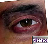
Symptoms include sudden onset of pain associated with bulbar movements, periorbital edema, erythema and swelling of the eyelids, proptosis, decreased visual acuity (if the optic nerve is involved) and diplopia ( in case of involvement of the extraocular muscles).
Treatment depends on the nature of the inflammatory reaction (nonspecific, granulomatous, or vasculitic) and may include administration of non-steroidal anti-inflammatory drugs, oral corticosteroids, radiotherapy, or immunomodulatory drugs. Recently, the use of monoclonal antibodies has also been introduced.
Orbital pseudotumor
Orbital pseudotumor (also called idiopathic inflammation of the orbit) is a nonspecific and idiopathic inflammation (it is not possible to identify a local or systemic cause). This process is characterized by the infiltration and proliferation of non-neoplastic cells in the mesenchymal tissues of the orbit. It is therefore a space-occupying lesion.
Typical symptoms of orbital pseudotumor include eye pain, eyelid redness and swelling, double vision, exophthalmos and decreased visual acuity.
In the most severe cases, the inflammation can cause a progressive fibrosis that leads to the so-called "frozen orbit", a real fixity of the eyeball characterized by ophthalmoplegia, ptosis and marked visual changes.
Important! The pseudotumor can simulate the symptoms of a neoplasm of the orbit. For this reason, the diagnostic tests must absolutely distinguish this pathology from the real tumors.
Orbital cellulite
Orbital cellulitis is an "infection of the orbital soft tissues, located behind the orbital septum. The disease is caused by the" extension of infectious processes by contiguity (nasal cavities, paranasal sinuses and dental elements), by the hematogenous spread of a "infection originating in an "other location or from the direct entry of pathogens following an orbital trauma that tears the orbital septum (eg animal bites, bruises or perforating lesions). The disease is characterized by an abrupt onset, with fever and a state of general malaise, associated with pain and reduced ocular mobility, conjunctival hyperemia and chemosis, redness and swelling of the eyelids and periorbital, visual clouding and proptosis. In many cases, signs of primary infection (eg nasal discharge and bleeding with sinusitis, periodontal pain and swelling with abscess etc.). Treatment must be timely and uses broad spectrum antibiotics and, in severe cases, surgery.
Preseptal cellulite
Preseptal and orbital (postseptal) cellulitis are two distinct diseases that share some clinical symptoms.
Preseptal cellulitis is an "infection of the eyelid and surrounding skin, located anterior to the orbital septum. This periorbital inflammation generally begins on the surface of the orbital septum, after the spread of secondary infections to local trauma of the face or eyelids. insect or animal bites, conjunctivitis, chalazion or sinusitis Both are particularly common in children, but preseptal cellulitis is far more frequent than orbital cellulitis.
Other inflammations of the orbit
- Dacryoadenitis: inflammatory process of the lacrimal glands, acute or chronic. Dacryoadenitis is common in children, following viral diseases such as measles and rubella. The chronic form is often associated with general diseases such as Sjogren's syndrome, sarcoidosis and Wegener's granulomatosis. Symptoms include fever, unilateral eyelid and periorbital pain and swelling; severe swelling can cause the eyeball to be displaced downwards and inwards. Treatment includes the use of antibiotics, anti-inflammatories and, in severe cases, immunosuppressive drugs.
- Myositis of the orbit: nonspecific inflammation of one or more extraocular muscles. It occurs at a young age, with ocular pain accentuated by the movements of the bulb and double vision. Often, it is associated with eyelid and periorbital edema, redness of the eye, ptosis and mild exophthalmos. Treatment involves the use of steroid anti-inflammatories and, in severe cases, immunosuppressive drugs.
- Toulouse-Hunt syndrome: idiopathic inflammation (ie of unknown origin) of the cavernous sinus, the superior orbital fissure and the orbital apex. It usually manifests itself with ocular pain accentuated by eye movements, double vision and ipsilateral headache. Hunt can also cause mild exophthalmos and oculomotor nerve paralysis. The disorder typically presents with acute phases alternating with periods of remission. Therapy involves the use of steroid anti-inflammatory drugs.
Orbital tumors
Orbital tumors can be primitive (ie they originate from the tissues of the orbit) or derive from neoplastic processes affecting contiguous structures (eyeball, ocular adnexa, paranasal sinuses and nasopharynx). Furthermore, the orbit can be affected by metastases.
Symptoms are variable, but typically an orbital expansive process produces eyeball protrusion (exophthalmos), eyelid ptosis, and double vision (diplopia). If optic nerve function is impaired, vision loss can result.
Orbital fractures
A violent trauma can cause the fracture of the bones of the facial massif. In many cases, this eventuality involves the involvement of various contiguous bone structures, such as the zygomatic-maxillary complex, the naso-orbito-ethmoid complex and the frontal sinus.
Due to their anatomical location and bone thickness, therefore, the orbital cavities are often involved, especially at the level of their lower wall (floor of the orbit). In these fractures, moreover, various other structures can be involved: the ocular musculature (rectus and inferior oblique muscle), the ocular globe, the optic and infraorbital nerve, the ophthalmic artery and vein.
Involvement of the orbital complex may be indicated by edema or periorbital ecchymosis, by anesthesia of the infraorbital nerve, by enophthalmos, by diplopia and by alterations in ocular motility. A lesion in the vicinity of the orbit always requires an eye examination, which includes at least the assessment of visual acuity, pupillary reactions and extraocular movements.

