Generality
Dermatomyositis is an idiopathic inflammatory disease that affects the muscles and skin, causing muscle deficits (weakness, pain and atrophy) and some typical skin signs (rash and scleroderma).

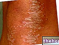
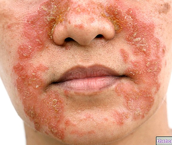
Figure: Skin signs associated with dermatomyositis. From the site: twicsy.com
In an advanced stage, dermatomyositis can also affect internal organs (esophagus, lungs and heart) and have serious consequences (difficulty swallowing, breathing problems and heart failure).
Currently, the causes of dermatomyositis are unknown, but an "immunological origin is hypothesized."
Diagnosis is based on a thorough physical examination, followed by several laboratory and instrumental tests.
The treatment methods currently available can only relieve symptoms and slow the progression of dermatomyositis.
What is dermatomyositis?
Dermatomyositis is a chronic inflammatory disease of connective tissues, characterized by skin (rash and scleroderma) and muscle (weakness, pain and atrophy) disorders. Not surprisingly, the name dermatomyositis derives from the "union of the terms" dermato ", which refers to the skin, and" myositis ", which refers to an" inflammation of the muscles.
If, in addition to the voluntary skeletal muscles, dermatomyositis also affects the striated muscles of the heart and the smooth muscles of the digestive, circulatory and respiratory systems, it can seriously endanger the life of affected people.
WHAT IS A MYOSITE?
Myositis is the medical term used to indicate a particular pathological condition, characterized by an "inflammation of the muscles of the body.
When a person suffers from myositis, the muscle fibers that make up his muscles deteriorate.
Depending on the triggering causes, myositis can be divided into:
- Idiopathic inflammatory myositis (N.B: in medicine, the term idiopathic means "without identifiable causes")
- Infectious myositis
- Myositis associated with other pathologies
- Myositis ossifying
- Drug-induced myositis
EPIDEMIOLOGY
According to a US statistical research, dermatomyositis has a frequency of 5-6 cases per million people. Therefore, it is an uncommon disease.
It can affect both adults and children: in adulthood, it usually appears around the age of 40-50, while in childhood / adolescence it usually occurs between 5 and 15 years.
For a reason that is still unclear, women get dermatomyositis significantly more than men.
Causes
The precise causes of origin of dermatomyositis are currently unknown.
Some researchers have tried to explain this disease as the result of a "viral (Epstein-Barr virus) or bacterial (Chlamydia pneumoniae And Chlamydia psittaci). Other scholars hypothesize that dermatomyositis is a pathological manifestation (therefore a symptom) of some autoimmune diseases, such as Sjögren's syndrome, systemic lupus erythematosus, rheumatoid arthritis or autoimmune vasculitis (NB: autoimmune diseases are particular conditions, in which the immune system of a person, instead of defending the latter from threats coming from outside, turns against him by attacking his organs).
Precisely because the causes are unknown, dermatomyositis is considered by doctors to be idiopathic inflammatory myositis.
Symptoms and Complications
The progressive deterioration of muscle fibers, which occurs as a result of dermatomyositis, is the cause of:
- Myalgia. It is pain in the muscles at the time of their contraction.
- Muscle asthenia. Synonymous with muscle weakness, it occurs mainly in the proximal voluntary musculature (affects the muscles that branch off directly from the trunk). The areas most affected are therefore the neck, shoulders, hips and thighs.
-
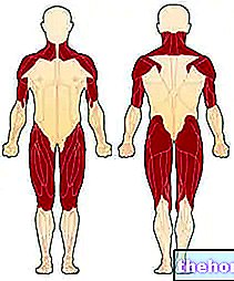
Figure: the first muscles affected by dermatomyositis. From the site: http://mda.org
Figure: the redness that accompanies dermatomyositis is characterized by uniform red-violet plaques. The rush tends to begin on the eyelids and then extend symmetrically to the face, arms, forearms and lower limbs. From the site: huidarts.com Muscle atrophy. It is the reduction of muscle mass (or tone). An atrophic musculature is less capable and less strong. Initially, muscle atrophy affects the muscles closest to the trunk (the same ones affected by asthenia); only later does it involve the distal musculature and that of the internal organs.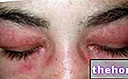
- Muscle soreness
As for the skin manifestations, the typical dermatomyositis rash involves the onset of red-purple spots in the eyelids, chest, face, back, hands and / or joints (knees and shoulders, in particular) .
The other characteristic sign of dermatomyositis, namely scleroderma, generally affects the arms and legs, but could also involve internal organs, such as kidneys, heart, esophagus, intestines and lungs. Scleroderma literally means "hard skin"; in fact this disease is characterized by an abnormal thickening of the skin, the result of an "excessive synthesis and deposition of collagen.
WHEN TO SEE THE DOCTOR?
The onset of muscle pain for no reason and the concomitant appearance of red-purple spots on the skin should prompt the person concerned to contact the doctor immediately for a clarification of the situation.
COMPLICATIONS
When muscle deterioration and scleroderma affect internal organs (esophagus, lungs, heart, etc.), the patient with dermatomyositis is life threatening, as he is subject to:
- Difficulty swallowing (dysphagia), followed by problems with nutrition and so-called pneumonia ab ingestis. These difficulties all arise from an "alteration of the smooth muscles of the digestive system (especially the first sections). The resulting nutritional problems lead to a sudden drop in body weight and the onset of a serious state of malnutrition.
N.B: pneumonia ab ingestis it is the inflammation of the lungs caused by the entry into the bronchial tree of food, saliva or nasal secretions. Its typical symptoms are: cough, fever, headache, dyspnoea and general malaise. - Respiratory problems. When the intercostal muscles that allow breathing are involved, and when scleroderma affects the respiratory tract, people with dermatomyositis breathe with enormous difficulty.
- Heart problems. Due to an "inflammation of the heart muscle (ie the myocardium), they can consist of arrhythmias of various kinds and heart failure.
In addition, especially among young patients, unusual accumulations of calcium in the skin and muscles (calcinosis) may occur.
ASSOCIATED DISEASES
Dermatomyositis can be associated with other disease states. In addition to the aforementioned autoimmune diseases, this pathology can be combined with:
- Raynaud's phenomenon. It is an excessive spasm of the peripheral blood vessels, which causes a reduction in blood flow to the involved regions.
The reaction can be triggered by cold and / or very intense emotional stress. The areas of the body most affected are the fingers and toes, the tip of the nose, the earlobes, the tongue and, in general, all those parts of the body crossed by small vessels and very susceptible to temperature variations.
Typical symptoms of Raynaud's phenomenon are: pain, burning, numbness and tingling. - Pulmonary interstitial disease. It is an alteration of the lining tissue of the pulmonary alveoli, or the cavities within which gas exchanges take place. In its most advanced stages, interstitial disease causes pulmonary fibrosis.
- Tumors in various organs of the body. In adults (especially of advanced age), dermatomyositis seems to favor the onset of tumors in the cervix, lungs, pancreas, breast, ovaries and gastrointestinal tract.
Diagnosis
Doctors use a physical examination and some laboratory and instrumental tests to determine whether certain signs and symptoms are attributable to dermatomyositis.
Among the various types of myositis, dermatomyositis is perhaps the simplest form to diagnose, as it combines muscle pain (which is common to many other diseases) with well-detailed signs on the skin.
OBJECTIVE EXAMINATION
During the physical examination, the doctor asks the patient to describe the symptoms felt and the exact location of the pain. After that, he dedicates himself to the observation of skin signs (rash) and to the palpation of painful muscles (NB: in case of dermatomyositis and myositis in general, the muscles are often soft and as if they had granules inside them). analyzes the patient's clinical history, investigating the possible presence of current and previous illnesses.
LABORATORY EXAMS
Laboratory tests consist of:
- Quantification of blood levels of creatine kinase, aldolase, autoantibodies and tumor antigens. Their dosage in a small blood sample is very useful for diagnostic purposes, because in an individual with dermatomyositis they are above normal. For example, creatine kinase is very high, even 50 times higher than normal (N.B: elevated creatine kinase indicates muscle damage).
- Skin biopsy. It consists in the collection and subsequent analysis, in the laboratory, of a small sample of skin cells from the area affected by the eruption.
This test is one of the most reliable methods for diagnosing dermatomyositis and for ruling out pathologies with similar symptoms. - Muscle biopsy. It consists in the collection and subsequent analysis, in the laboratory, of a small sample of muscle cells from the painful areas.
While it is useful for detecting muscle damage or infection, it is less reliable than a skin biopsy.
INSTRUMENTAL EXAMINATIONS
The possible instrumental tests are:
- Electromyography. It is used to measure the electrical activity of the muscles. It is not invasive at all.
- Nuclear magnetic resonance (NMR). By creating magnetic fields, MRI provides a "detailed image of the muscles. It is not an invasive exam."
- Chest X-ray. It is used to assess the health of the lungs. It is important when there is a suspicion of lung involvement. It is considered an invasive test because it exposes the patient to a minimal dose of ionizing radiation.
Treatment
At present, there is still no specific cure for dermatomyositis.
Current treatments available to patients can only improve the symptomatological picture (including complications) and slow down the progression of the disease.
Among the various treatments, corticosteroid and immunosuppressive drugs play a primary role, as well as rehabilitation treatments and surgery.
According to some scientific studies, the sooner symptomatic therapy begins, the greater the benefits the patient will enjoy.
PHARMACOLOGICAL THERAPY
As anticipated, the most used drugs in case of dermatomyositis are corticosteroids and immunosuppressants.
The former are powerful anti-inflammatory drugs, while the latter are responsible for lowering the immune defenses. Both are administered with the ultimate aim of reducing inflammation and the autoimmune response (NB: corticosteroids are also useful in maintaining muscle strength and in "avoiding" atrophy of the muscles subject to deterioration).
If the aforementioned drugs prove ineffective, doctors may resort to a third possibility, represented by intravenous immunoglobulins. These, like previous drugs, work by reducing the autoimmune response, but are much more expensive.
What are the side effects of corticosteroids and immunosuppressants?
If taken for long periods and / or in high doses, corticosteroids can cause serious side effects, such as diabetes, osteoporosis, hypertension, weight gain, cataracts etc.
Immunosuppressants, on the other hand, make the subject who uses them more fragile and more exposed to infections.
- Prednisone
- Methylprednisolone
- Topical
- Systemic
- Methotrexate
- Azathioprine
- Rituximab
- Cyclophosphamide
- Mycophenolate mofetil
- Cyclosporine
- Tacrolimus
- Infliximab
- Systemic
- Intravenous
REHABILITATION THERAPY
Depending on the severity of symptoms, patients with dermatomyositis may need to undergo:
- Physiotherapy. Thanks to the help of an expert physiotherapist, the patient can maintain a moderate muscle tone, even in spite of the progressive deterioration that the muscles undergo. It is essential that patients learn to perform motor exercises in full autonomy, in such a way as to to be able to play them at home, in their free time.
- Speech therapy. It is indicated for patients with swallowing problems, as it helps to reduce its severity.
- Adequate diet. Those who have problems eating, it is good that they know which foods to eat for a correct nutritional intake. Therefore, a dietician will take care of preparing a diet commensurate with the age and needs of the patient.
SURGERY
The only surgical treatment that can be used in the case of dermatomyositis is to remove calcium deposits in the skin and muscles (calcinosis).
SOME ADVICES
Dermatomyositis weakens the skin, especially the areas affected by the rash.
Therefore, doctors recommend not exposing yourself too much to the sun or doing it only after taking the necessary precautions (protective sunscreen, suitable clothing, etc.), as ultraviolet rays could further worsen the situation.
Furthermore, it is a good idea:
- Contact the attending physician for any information regarding dermatomyositis and possible associated pathologies.
- Stay active, in such a way as to tone the muscles of the body.
- In case of extreme fatigue, rest and allow your body to recover strength.
- If you have any signs or symptoms you have never experienced before, contact your doctor immediately.
- If you are depressed due to illness, confide in friends and family. In fact, closing in on oneself does not help the therapies.
Prognosis
At one time, when current treatments did not yet exist, the prognosis was negative and the 5-year survival rate from onset of symptoms was very low.
Today, however, thanks to medical advances, the prognosis has improved a lot and the survival rates at 5 and 10 years from the onset of dermatomyositis are respectively 70% and 57%.




.jpg)


