A repeated irritative phenomenon, a trauma, an infection or a genetic mutation can determine the appearance of an "exostosis"; in a non negligible number of cases, however, these benign new formations of bone tissue are due to unknown reasons.
There are numerous types of exostoses, including: auditory canal exostosis, buccal exostosis, heel spur, multiple hereditary exostosis, osteochondroma, sinus osteoma and sub-nail exostosis.
Exostoses can be symptomatic or asymptomatic; when they are symptomatic, the associated manifestations depend on the anatomical site involved.
Diagnosed by X-rays, exostoses require treatment when they are responsible for symptoms that affect the patient's quality of life.
The word exostosis, therefore, includes all the possible bone growths of a benign nature that can be generated on the surface of a bone, including the so-called osteophytes (or bone spurs) and osteochondromas.
- Osteophyte is the name of the bone formation similar to a claw or a rose thorn, which can form near the joints, as a result of chronic irritation or long-lasting erosive processes.
- Osteochondroma, on the other hand, is the medical term that defines exostoses with an appearance on the cartilage portion of those bones that include a layer of cartilage.
Exostoses: the most common sites
All bones in the human body can be subjected to exostosis.
However, there are bones which, due to their localization or for reasons not yet elucidated, are more affected than others; more specifically, among the bones most subject to the phenomenon of "exostosis, there are:
- The bones that make up the external auditory canal;
- The bones of the ankle;
- The calcaneus, one of the 7 bones of the tarsus of the foot;
- The mandible or maxilla;
- The long bones of the limbs (humerus, radius, ulna, phalanges of the hands, femur, tibia and fibula);
- The bones make up the paranasal sinuses (sphenoid, ethmoid, etc.).
The cases of exostosis due to a "DNA alteration (no matter if hereditary or acquired during embryonic development) take the generic name of multiple hereditary exostosis;
It should be noted that, in spite of what has just been reported, many cases of exostosis appear for unknown causes (i.e. their appearance cannot be attributed to a specific phenomenon or episode).
Most common types of exostoses
The most common and described types of exostoses are:
- The exostosis of the ear canal;
- The heel spur;
- The multiple hereditary exostosis;
- The osteoma of the paranasal sinuses;
- The buccal exostosis;
- The sub-nail exostosis;
- The osteochondroma.
EXOSTOSES OF THE HEARING CONDUCT

Also known as surfer's ear, exostosis of the ear canal is the condition resulting from the growth of a bony growth on the surface of the bones that form the external auditory canal (it is the canal of the external ear, which begins at the level of the auricle and leads to the eardrum).
Currently, doctors have not yet identified the precise cause of the ear canal exostosis; however, having detected a "high incidence of this condition in people practicing water sports such as surfing, sailing, etc., they believe it is a determining causal factor. the repeated exposure of the external auditory canal to water and wind (NB: this also explains the expression "surfer's ear").
Ear canal exostosis can affect one or both ears and can degenerate into hearing loss.
CALCANEAN SPINE

Shutterstock
Also known as a heel spur, the heel spur is the exostosis of the heel.
Similar to a claw or a rose thorn, the heel spur is usually the result of the continuous repetition of phenomena that irritate the insertion of the Achilles tendon on the heel (in this case, the exostosis will be located on the posterior front. of the foot) or the insertion of the plantar fascia on the heel (in this situation, however, the exostosis will be localized on the inferior-posterior portion of the foot).
Statistics say that heel spurs are more common in:
- People who have the habit of wearing shoes that "hit" the back of the heel or that significantly modify the arch (eg shoes with heels, in the case of women);
- People who practice sports such as road running, in which it is possible to develop an "inflammation of the plantar fascia (plantar fasciitis);
- Individuals who by their nature have an Achilles tendon that is narrower than normal;
- Obese or overweight subjects.
MULTIPLE HEREDITARY EXOSTOSIS
The aforementioned multiple hereditary exostosis is a genetic disease, which results in the formation of different bone growths in various bones of the human body.
Hereditary condition in 50% of cases and acquired in the course of embryonic development in the remaining percentage, the multiple hereditary exostosis preferably affects the long bones of the leg, shoulders and shoulder blades.
Multiple hereditary exostosis tends to go unnoticed until the age of 5-6, when the patient begins to develop the first abnormal bone growths.
OSTEOMA OF THE PARANASAL SINUS
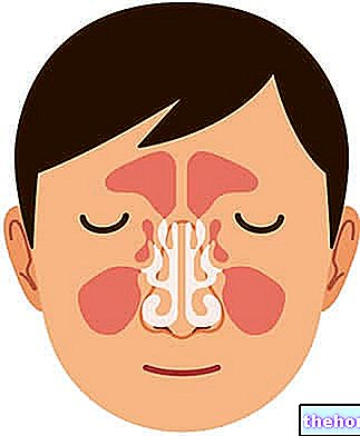
The paranasal sinuses are the 4 air-filled cavities located inside the cheeks and forehead, and resulting from the particular arrangement of the ethmoid cranial bones (ethmoid sinuses), sphenoid (sphenoid sinuses), frontal (frontal sinuses) and maxillary ( maxillary sinuses).
The paranasal sinuses are used to: improve the perception of odors, amplify the sounds and voice emitted through the vocal cords, make the skull less heavy and humidify-warm-purify the inhaled air.
The osteoma of the paranasal sinuses is the condition that can arise as a result of an exostosis of one of the ethmoid bone, the sphenoid bone, the frontal bone and the maxillary bone.
Due to completely unknown causes, the osteoma of the paranasal sinuses can be an obstacle to the passage of inhaled air and the drainage of mucus.
BUCCAL EXOSTOSES
Buccal exostosis is the medical expression that defines the benign bone formations that can be generated inside the mouth, or on the jaw or mandible.
As a rule, at the "origin of the episodes of buccal exostosis c" is a trauma or an injury to the gum (with obviously an involvement of the underlying bone structure).
Buccal exostosis is more common in adolescence.
SUB-NAUGH EXOSTOSIS
Also known as nail bed exostosis, sub-nail exostosis is the condition resulting from the formation of an abnormal outgrowth on the bony surface immediately below the so-called nail bed (ie the characteristic portion of the fingers and toes, on which the "nail).
Generally, at the origin of the episodes of sub-nail exostosis c is a trauma to the finger tract that develops the anomaly.
Statistics in hand, sub-nail exostosis is a phenomenon that doctors most often find in the first toe, that is the big toe.
Teenagers suffer most from nail bed exostosis.
OSTEOCONDROME
As already exposed to the readers, an osteochondroma is an "exostosis that forms on the cartilage surface of a bone, which means that" osteochondroma is an abnormal bone growth, covered, unlike normal exostoses, by a layer of cartilage.
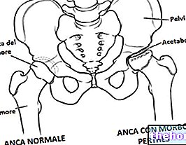
Also known as osteo-cartilage exostosis, osteochondroma most often affects the bones of the lower limbs, the bones of the pelvis (particularly those involved in the hip joint) and the scapula.
Among the various types of exostoses recognized and described in the medical literature, osteochondroma is the common one; in this regard, epidemiological investigations report that about 2% of the general population suffer from osteochondroma.
Currently, the causes of osteochondromas are unknown; however, experts believe that the formation of these osteo-cartilaginous growths is affected by some anomaly of skeletal development, since the most affected subjects are children and adolescents (ie a category of person in full phase of bone growth).
Complications
Although they are benign formations, exostoses can still be at the origin of complications. For example:
- The exostosis of the auditory canal can cause complications such as: hearing loss and tendency to develop recurrent ear infections (they are due to water that accumulates and stagnates in the ear canal, following the altered anatomy of the latter) ;
- If it grows close to blood vessels, osteochondroma can lead to complications of a vascular nature, including: false aneurysm (or pseudoaneurysm), phlebitis and acute ischemia.
- Multiple hereditary exostosis can become complicated when one of the bone growths becomes malignant (in these situations, the resulting malignant tumor is an example of osteosarcoma).
Did you know that ...
According to statistics, the evolution in malignant terms of a "bone growth due to" multiple hereditary exostosis would affect between 1 and 6 patients out of 100, fortunately, therefore, it is an uncommon phenomenon.
.For subjects suffering from multiple hereditary exostoses, the diagnosis of the genetic condition in question is possible even before birth (NB: doctors search for this disease only when one of the parents of the possible future patient is a carrier; otherwise , it is not a routine test, as could be, for example, the one related to Down syndrome).
or a helmet that protects the ears from exposure to water and wind.
CALCANEAR SPINE
For the treatment of symptomatic cases of heel spurs, there are two types of therapeutic approach: a therapeutic approach of a conservative nature, which represents the first line treatment, and a therapeutic approach of a surgical nature, which is instead the treatment reserved exclusively for patients on which the aforementioned conservative therapies have been ineffective.
- The conservative treatment of the heel spur includes: rest from all those activities that could cause heel pain, intake of anti-inflammatories, application of ice on the painful area, exercises of stretching and strengthening of the leg muscles (obviously of the lower limb affected by the exostosis), physiotherapy and use of shoes that preserve the health of the heel (for a woman, the use of heeled shoes is to be avoided);
- The surgical treatment of the heel spur, on the other hand, consists in the "removal of the exostosis", followed by a period of rest and physiotherapeutic rehabilitation.
MULTIPLE HEREDITARY EXOSTOSIS
When it is a source of symptoms, multiple hereditary exostosis requires the use of surgical therapy aimed at:
- Remove all exostoses that compress neighboring nerves and / or blood vessels;
- Remove any exostoses that show signs of malignancy;
- Correct deviations of the limbs;
- Correct, as far as possible, the heterometry of the limbs.

